. 2024 Jan 23:11:1232269.
doi: 10.3389/fcvm.2024.1232269. eCollection 2024.

Combinatorial immune checkpoint blockade increases myocardial expression of NLRP-3 and secretion of H-FABP, NT-Pro-BNP, interleukin-1β and interleukin-6: biochemical implications in cardio-immuno-oncology

Affiliations

Combinatorial immune checkpoint blockade increases myocardial expression of NLRP-3 and secretion of H-FABP, NT-Pro-BNP, interleukin-1β and interleukin-6: biochemical implications in cardio-immuno-oncology

V Quagliariello et al. Front Cardiovasc Med. .

Abstract

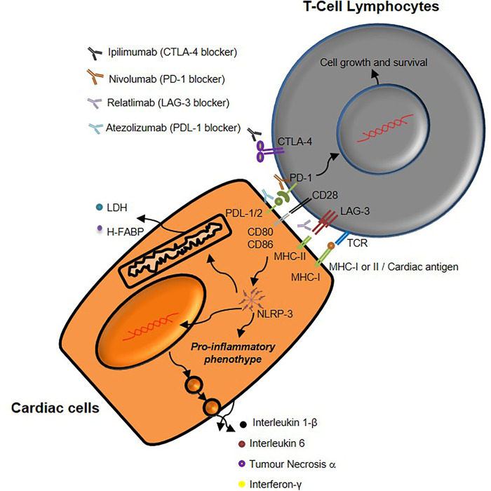
Background: Immune checkpoint blockade in monotherapy or combinatorial regimens with chemotherapy or radiotherapy have become an integral part of oncology in recent years. Monoclonal antibodies against CTLA-4 or PD-1 or PDL-1 are the most studied ICIs in randomized clinical trials, however, more recently, an anti-LAG3 (Lymphocyte activation gene-3) antibody, Relatlimab, has been approved by FDA in combination with Nivolumab for metastatic melanoma therapy. Moreover, Atezolizumab is actually under study in association with Ipilimumab for therapy of metastatic lung cancer. Myocarditis, vasculitis and endothelitis are rarely observed in these patients on monotherapy, however new combination therapies could expose patients to more adverse cardiovascular events.

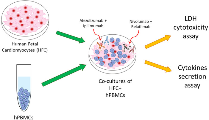
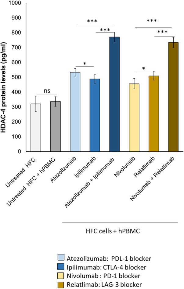
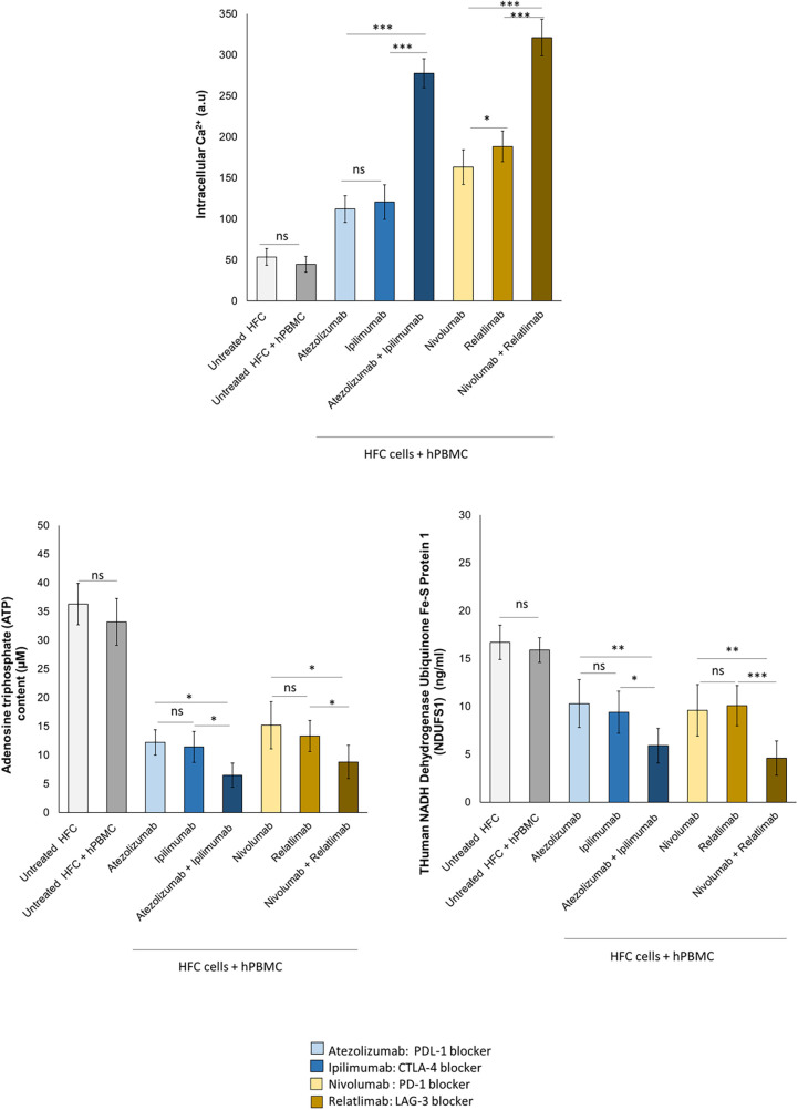
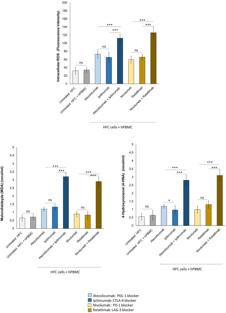
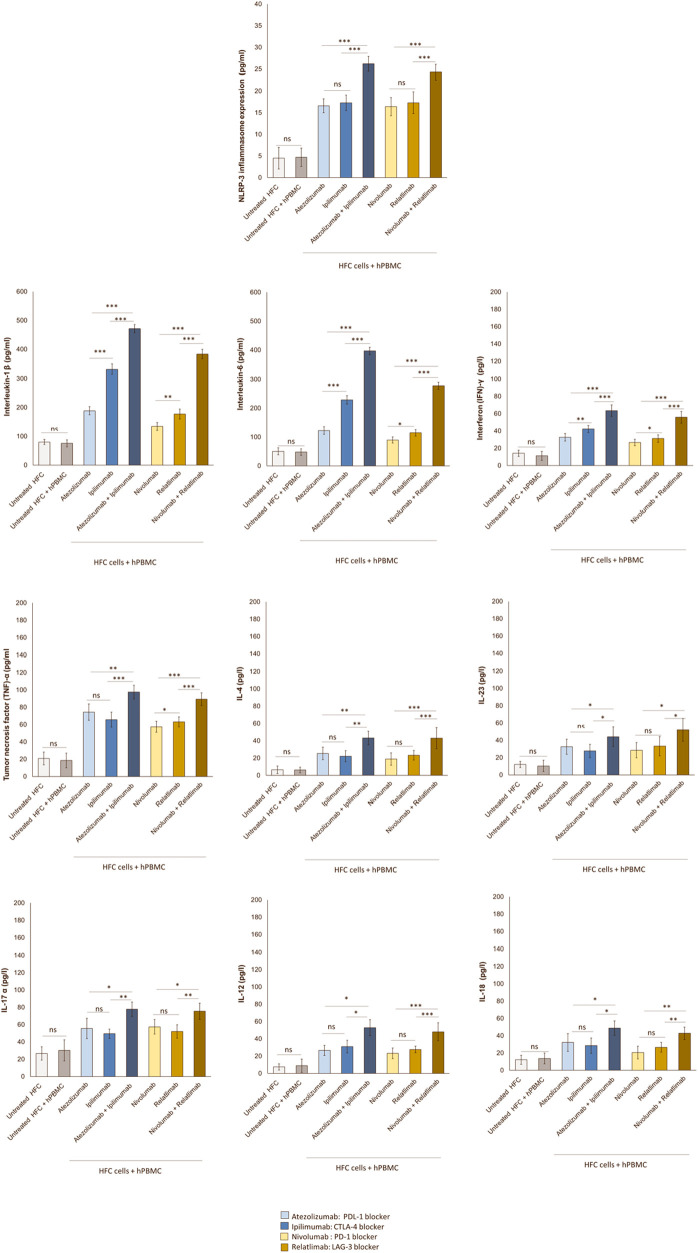
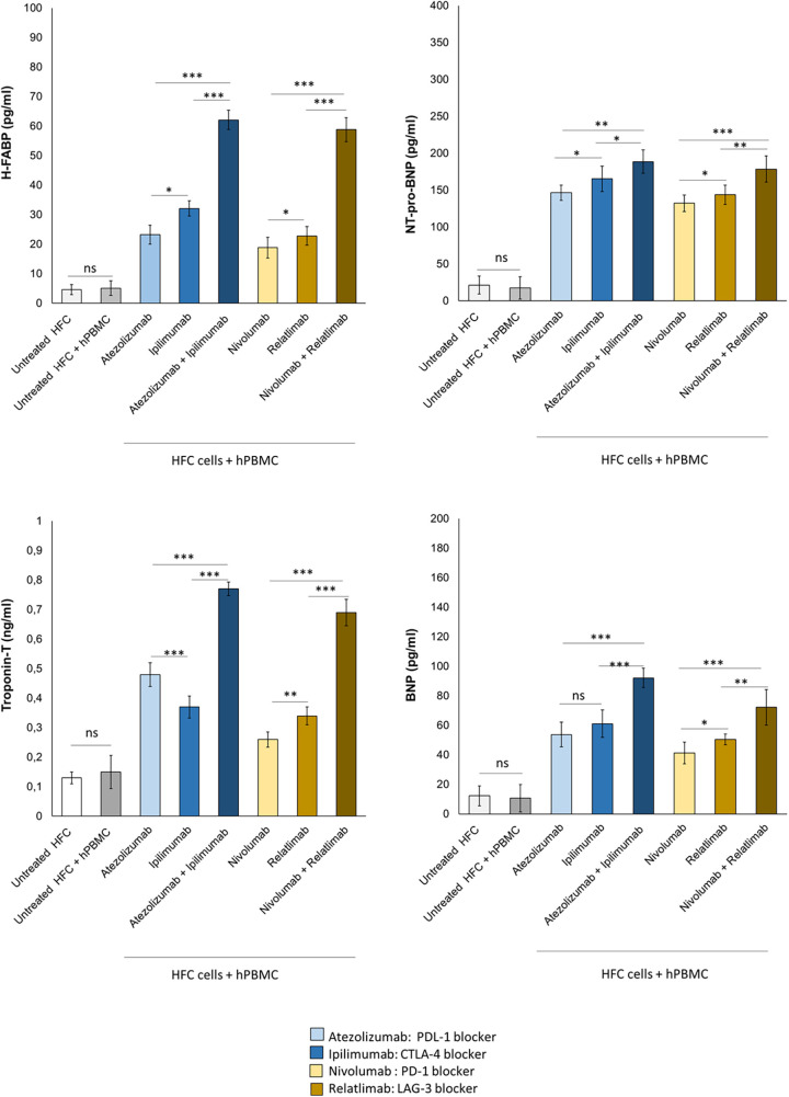
Methods: Human cardiomyocytes co-cultured with human peripheral blood lymphocytes (hPBMCs) were exposed to monotherapy and combinatorial ICIs (PD-L1 and CTLA-4 or PD-1 and LAG-3 blocking agents, at 100 nM) for 48 h. After treatments, cardiac cell lysis and secretion of biomarkers of cardiotoxicity (H-FABP, troponin-T, BNP, NT-Pro-BNP), NLRP3-inflammasome and Interleukin 1 and 6 were determined through colorimetric and enzymatic assays. Mitochondrial functions were studied in cardiomyocyte cell lysates through quantification of intracellular Ca++, ATP content and NADH:ubiquinone oxidoreductase core subunit S1 (Ndufs1) levels. Histone deacetylases type 4 (HDAC-4) protein levels were also determined in cardiomyocyte cell lysates to study potential epigenetic changes induced by immunotherapy regimens.

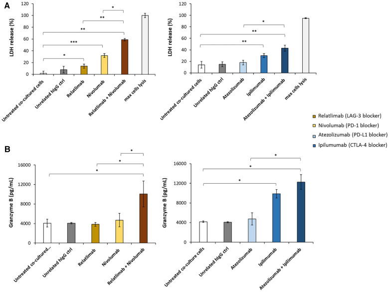
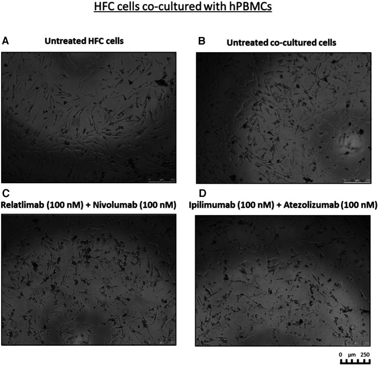
Results: Both combinations of immune checkpoint inhibitors exert more potent cardiotoxic side effects compared to monotherapies against human cardiac cells co-cultured with human lymphocytes. LDH release from cardiac cells was 43% higher in PD-L1/CTLA-4 blocking agents, and 35.7% higher in PD-1/LAG-3 blocking agents compared to monotherapies. HDAC4 and intracellular Ca++ levels were increased, instead ATP content and Ndufs1 were reduced in myocardial cell lysates (p < 0.001 vs. untreated cells). Troponin-T, BNP, NT-Pro-BNP and H-FABP, were also strongly increased in combination therapy compared to monotherapy regimen. NLRP3 expression, IL-6 and IL-1β levels were also increased by PDL-1/CTLA-4 and PD-1/LAG-3 combined blocking agents compared to untreated cells and monotherapies.

Conclusions: Data of the present study, although in vitro, indicate that combinatorial immune checkpoint blockade, induce a pro- inflammatory phenotype, thus indicating that these therapies should be closely monitored by the multidisciplinary team consisting of oncologists, cardiologists and immunologists.

Keywords: cancer; cardiology; cardiotoxicity; immune checkpoint inhibitors; inflammation; myocarditis; oncology.


Conflict of interest statement

All authors declare that the research was conducted in the absence of any commercial or financial relationships that could be construed as a potential conflict of interest.

Figures

Figure 1
Figure 1
Schematical illustration of HFC + hPBMCs co-cultures as model of ICIs-mediated cardiotoxicity due to direct interaction between cardiac and immune cells.
Figure 2
Figure 2
Analysis of LDH and granzyme levels released by co-cultures of HFC cells with hPBMCs. HFC cells were incubated with or without hPBMC and treated for 48 h with Relatlimab or Nivolumab (left panel) and with Atezolizumab or Ipilimumab (right panel), used at the concentration of 100 nM as single agents or in combination. (A) The cytotoxic effects of the mAbs on HFC cells was analyzed by measuring the levels of LDH release in the supernatants of the co-cultured cells, following the manufacturing recommendations of the CyQUANT LDH Cytotoxicity Assay by ThermoFisher. (B) The levels of Granzyme B were measured in the supernatants of co-cultures by using the ELISA Granzyme B kit, as described in methods. A human unrelated IgG was used as control. The values reported are related to the mean of three experiments ± SD); p-values were calculated by comparing each treatment with untreated co-cultured cells or the combined treatment with each single treatment and reported as ***p < 0.001; **p < 0.01; *p < 0.05.
Figure 3
Figure 3
Cell debris and significant reduction in fibroblast-like phenotype of HFC cells, co-cultured with hPBMCs, exposed to combinatorial ICIs treatments. Microscopy Images of the co-cultures after treatment with combinations of Relatlimab and Nivolumab (C) or Ipilimumab and Atezolizumab (D), respectively, at the indicated concentrations. Untreated HFC cells in the absence (A) or in the presence of hPBMCs (B) were used in parallel as negative controls. The images were obtained via Leica Advanced microscopy (Leica DMI4000 B).
Figure 4
Figure 4
ICIs therapies increased HDAC-4 expression in HFC cells co-cultured with hPBMCs. HFC cells were incubated with or without hPBMC and treated for 48 h with Atezolizumab or Ipilimumab or Relatlimab or Nivolumab (100 nM). HDAC-4 expression (pg/ml), in HFC lysate, were quantified through selective ELISA kit, as described in methods. The data represent the mean ± SD of three independent experiments ***p < 0.001; **p < 0.01; *p < 0.05.
Figure 5
Figure 5
ICIs therapies increased intracellular Ca++ levels, decreased ATP production and NDUFS1 levels in HFC cells co-cultured with hPBMCs. HFC cells were incubated with or without hPBMC and treated for 48 h with Atezolizumab or Ipilimumab or Relatlimab or Nivolumab (100 nM). Intracellular Ca++ levels (a.u), in HFC lysate, were quantified through fluorescent Kit, as described in methods. Intracellular ATP (µM) and NDUFS1 (ng/ml) levels were quantified through ELISA methods, as described in methods. The data represent the mean ± SD of three independent experiments ***p < 0.001; **p < 0.01; *p < 0.05. Differences between control group (Untreated HFC + hPBMC) and ICIs groups were always statistically significant (p < 0.001 for all).
Figure 6
Figure 6
ICIs therapies increased intracellular ROS and lipid peroxidation products (MDA and 4-HNA) levels in HFC cells co-cultured with hPBMCs. HFC cells were incubated with or without hPBMC and treated for 48 h with Atezolizumab or Ipilimumab or Relatlimab or Nivolumab (100 nM). Intracellular ROS levels (fluorescence intensity), in HFC lysate, were quantified through fluorescent Kit, as described in methods. Intracellular lipid peroxidation products, such as MDA and 4-HNA (mmol/ml) levels were quantified through colorimetric assay, as described in methods. The data represent the mean ± SD of three independent experiments ***p < 0.001; **p < 0.01; *p < 0.05.
Figure 7
Figure 7
Combinatorial ICIs therapies increases NLRP3 expression and release of several pro-inflammatory biomarkers in co-cultures of HFC cells with hPBMCs. HFC cells were incubated with or without hPBMC and treated for 48 h with Atezolizumab or Ipilimumab or Relatlimab or Nivolumab (100 nM). NLRP-3 inflammasome expression (in HFC lysate), IL-1β, IL-6, TNF-α, IL-4, IL-23, IL-17a, IL-12, IL-18 and INF-γ release in surnatant (pg/ml) were quantified through selective ELISA kits, as described in methods. The data represent the mean ± SD of three independent experiments ***p < 0.001; **p < 0.01; *p < 0.05.
Figure 8
Figure 8
Combinatorial ICIs therapies increase cardiotoxicity biomarkers in co-cultures of HFC cells with hPBMCs. HFC cells were incubated in the absence or in the presence of human lymphocytes and treated for 48 h with Atezolizumab or Ipilimumab or Relatlimab or Nivolumab at 100 nM as single agents or in combination. H-FABP expression (pg/ml), NT-pro-BNP (pg/ml), Troponin T (ng/ml)and BNP (pg/ml) expression were quantified in co-cultures through selective ELISA kits, as described in methods. The data represent the mean ± SD of three independent experiments ***p < 0.001; **p < 0.01; *p < 0.05.
Figure 9
Figure 9
Schematic representation of mechanisms underlying immune-related cardiac toxicity due to CTLA-4, PD-1 and LAG-3 blocking agents therapy.

References

    1. España Fernandez S, Sun C, Solé-Blanch C, Boada A, Martínez-Cardús A, Manzano JL. Immunotherapy resumption/rechallenge in melanoma patients after toxicity: do we have another chance? Pharmaceutics. (2023) 15(3):823. 10.3390/pharmaceutics15030823 - DOI - PMC - PubMed
    1. Wang S, Li Y, Liu Z, Tian W, Zeng Y, Liu J, et al. Efficacy and safety of first-line immune checkpoint inhibitors combined with chemotherapy for extensive-stage small cell lung cancer: a network meta-analysis. Lung Cancer. (2023) 178:47–56. 10.1016/j.lungcan.2023.02.003 - DOI - PubMed
    1. Hellmann MD, Friedman CF, Wolchok JD. Combinatorial cancer immunotherapies. Adv Immunol. (2016) 130:251–77. 10.1016/bs.ai.2015.12.005 - DOI - PubMed
    1. Vilgelm AE, Johnson DB, Richmond A. Combinatorial approach to cancer immunotherapy: strength in numbers. J Leukoc Biol. (2016) 100(2):275–90. 10.1189/jlb.5RI0116-013RR - DOI - PMC - PubMed
    1. Goldberg MV, Drake CG. LAG-3 in cancer immunotherapy. Curr Top Microbiol Immunol. (2011) 344:269–78. 10.1007/82_2010_114 - DOI - PMC - PubMed