. 2024 Jan 19;27(2):108969.
doi: 10.1016/j.isci.2024.108969. eCollection 2024 Feb 16.

The GDNF-gel/HA-Mg conduit promotes the repair of peripheral nerve defects by regulating PPAR-γ/RhoA/ROCK signaling pathway

Affiliations

The GDNF-gel/HA-Mg conduit promotes the repair of peripheral nerve defects by regulating PPAR-γ/RhoA/ROCK signaling pathway

Yuanqing Cai et al. iScience. .

Abstract

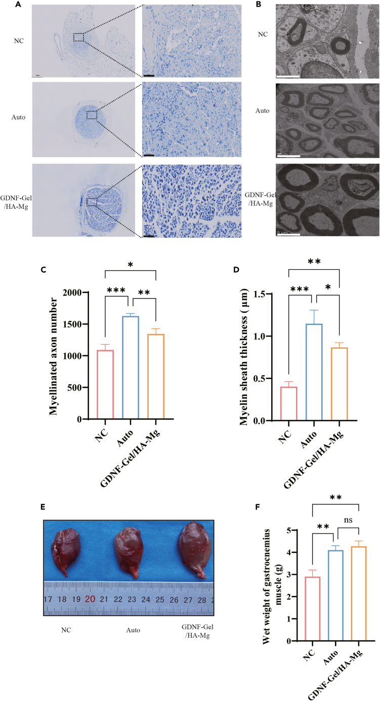
Magnesium (Mg)-based conduits have gained more attention in repairing peripheral nerve defects. However, they are limited due to poor corrosion resistance and rapid degradation rate. To tackle this issue, glial cell line-derived neurotrophic factor (GDNF)- Gelatin methacryloyl (Gel)/hydroxylapatite (HA)-Mg nerve conduit was developed and implanted in sciatic nerve defect model in Sprague-Dawley (SD) rats. The sciatic functional index measurement showed that the GDNF-Gel/HA-Mg nerve conduit effectively promoted the recovery of sciatic nerve function. The pathological examination results showed that there were more regenerated nerve tissues in GDNF-Gel/HA-Mg group, with a higher number of regenerating axons, and the thickness of the myelin sheath was significantly larger than that of control group (NC group). Immunofluorescence results revealed that the GDNF-Gel/HA-Mg conduit significantly promoted the expression of genes associated with nerve repair. RNA-seq and molecular test results indicated that GDNF-Gel/HA-Mg might be involved in the repair of peripheral nerve defects by regulating PPAR-γ/RhoA/ROCK signaling pathway. Biological sciences; Neuroscience; Molecular neuroscience; Techniques in neuroscience.

Keywords: Biological sciences; Molecular neuroscience; Neuroscience; Techniques in neuroscience.


Conflict of interest statement

The authors declare that they have no competing interests.

Figures

None
Graphical abstract
Figure 1
Figure 1
Graphic abstract: the preparation of GDNF-Gel/HA-Mg conduit and its potential value in repairing peripheral nerve defect Mg, magnesium; HA, hydroxylapatite; GDNF, glial cell line-derived neurotrophic factor; GelMA, Gelatin methacryloyl; PPAR-γ, peroxisome proliferator-activated receptor gamma; ROCK, Rho-associated kinase.
Figure 2
Figure 2
The effect of GDNF-Gel/HA-Mg conduit on sciatic functional index and never tissues regeneration (A and B) diagram for implantation and suture of GDNF-Gel/HA-Mg conduit in vivo. (C and D) the measurement of sciatic functional index and statistical analysis at 12 weeks post-surgery. (E and F) Hematoxylin and Eosin (HE) staining of cross-sectional and longitudinal sections of regenerated nerves harvested at 12 weeks post-surgery. NC, negative control; Auto, autograft; GDNF, glial cell line-derived neurotrophic factor; GelMA, Gelatin methacryloyl; HA, hydroxylapatite; Mg, magnesium; SFI, sciatic functional index; ns, no statistical difference; ∗p < 0.05; ∗∗p < 0.01; ∗∗∗p < 0.001. scale bar: E:100μm; F: 50μm. Data are represented as mean ± SEM.
Figure 3
Figure 3
The effect of GDNF-Gel/HA-Mg conduit on never tissues regeneration and neuro-electrophysiology (A and D) Masson Staining of regenerated nerves harvested at 12 weeks post-surgery and statistical analysis. (B) Sirius Red Staining of regenerated nerves harvested at 12 weeks post-surgery. (C) Assessment of the extent of adherence between regenerated nerves and adjacent tissues. (E and F) The nerve conduction velocity and latency measurement of regenerated nerves at 12 weeks post-surgery. NC, negative control; Auto, autograft; GDNF, glial cell line-derived neurotrophic factor; GelMA, Gelatin methacryloyl; HA, hydroxylapatite; Mg, magnesium; ns, no statistical difference; ∗p < 0.05; ∗∗p < 0.01; ∗∗∗p < 0.001. scale bar: A:100μm; B: 50μm. Data are represented as mean ± SEM.
Figure 4
Figure 4
The effect of GDNF-Gel/HA-Mg conduit on regenerated axons and myelin (A and C) Toluidine blue (TB) staining of regenerated nerves harvested at 12 weeks post-surgery and statistical analysis. (B and D) myelin sheath thickness detected by transmission electron microscopy (TEM) and statistical analysis. (E and F) Gastrocnemius muscle wet weight measurement and statistical analysis at 12 weeks post-surgery. NC, negative control; Auto, autograft; GDNF, glial cell line-derived neurotrophic factor; GelMA, Gelatin methacryloyl; HA, hydroxylapatite; Mg, magnesium; ns, no statistical difference; ∗p < 0.05; ∗∗p < 0.01; ∗∗∗p < 0.001. scale bar: A:50μm; B: 5μm. Data are represented as mean ± SEM.
Figure 5
Figure 5
The expression of S100 and p75 at 12 weeks post-surgery (A and C) The expression of S100 detected by immunofluorescence and statistical analysis. (B and D) The expression of p75 detected by immunofluorescence and statistical analysis.NC, negative control; Auto, autograft; GDNF, glial cell line-derived neurotrophic factor; GelMA, Gelatin methacryloyl; HA, hydroxylapatite; Mg, magnesium; SFI, sciatic functional index; ns, no statistical difference; ∗∗p < 0.01; ∗∗∗p < 0.001. scale bar: 50μm. Data are represented as mean ± SEM.
Figure 6
Figure 6
The expression of neurofilament-200 (NF-200) and myelin basic protein (MBP) at 12 weeks post-surgery (A) The expression of neurofilament-200 (NF-200) and myelin basic protein (MBP) detected by immunofluorescence. (B) statistical analysis. NF-200, neurofilament-200; MBP, myelin basic protein; NC, negative control; Auto, autograft; GDNF, glial cell line-derived neurotrophic factor; GelMA, Gelatin methacryloyl; HA, hydroxylapatite; Mg, magnesium; SFI, sciatic functional index; ns, no statistical difference; ∗∗∗p < 0.001. scale bar: 50μm. Data are represented as mean ± SEM.
Figure 7
Figure 7
The bioinformatics analysis results (A and B) Principal Component Analysis (PCA) and Cluster analysis of samples. (C) Volcano map of differentially expressed genes. (D) The mRNA expression level of differentially expressed gene. (E) Cluster analysis of differentially expressed genes. (F) GO Enrichment Analysis of differentially expressed genes. (G) KEGG Pathway Enrichment Analysis of differentially expressed genes. (H) Gene Set Enrichment Analysis (GSEA) of PPAR signaling pathway between NC and GDNF-Gel/HA-Mg group; NC, negative control; GDNF, glial cell line-derived neurotrophic factor; GelMA, Gelatin methacryloyl; HA, hydroxylapatite; Mg, magnesium; FC, fold change.
Figure 8
Figure 8
The expression levels of PPAR-γ/RhoA/ROCK signaling pathway (A and B) The expression of PPAR-γ detected by immunofluorescence and statistical analysis; (C‒E) the relative mRNA expression level of PPAR-γ, Rho, ROCK and statistical analysis; (F‒I) The expression of peroxisome proliferator-activated receptor gamma (PPAR-γ), RhoA, Rho-associated kinase (ROCK) by western blotting and statistical analysis. NC: negative control; GDNF: glial cell line-derived neurotrophic factor; GelMA: Gelatin methacryloyl; HA: hydroxylapatite; Mg: magnesium; PPAR-γ: peroxisome proliferator-activated receptor gamma; ROCK: Rho-associated kinase; ∗p < 0.05; ∗∗p < 0.01. scale bar: 50μm. Data are represented as mean ± SEM.
Figure 9
Figure 9
Biological safety assessment of GDNF-Gel/HA-Mg in vivo (A) The levels of myocardial enzymes, transaminases, and renal function and quantitative analysis. (B‒F) HE results of heart (B), liver(C), spleen(D), lung(E) and kidney(F) from different groups. CK, creatine kinase; CK-MB, creatine kinase isoenzyme-MB; LDH, lactic dehydrogenase; ALT, alanine aminotransferase; AST, aspartate aminotransferase; NC, negative control; GDNF, glial cell line-derived neurotrophic factor; GelMA, Gelatin methacryloyl; HA, hydroxylapatite; Mg, magnesium; ns, no statistical difference. Scale bar: 100μm. Data are represented as mean ± SEM.

References

    1. Modrak M., Talukder M.A.H., Gurgenashvili K., Noble M., Elfar J.C. Peripheral nerve injury and myelination: Potential therapeutic strategies. J. Neurosci. Res. 2020;98:780–795. doi: 10.1002/jnr.24538. - DOI - PMC - PubMed
    1. Wang M.L., Rivlin M., Graham J.G., Beredjiklian P.K. Peripheral nerve injury, scarring, and recovery. Connect. Tissue Res. 2019;60:3–9. doi: 10.1080/03008207.2018.1489381. - DOI - PubMed
    1. Battiston B., Titolo P., Ciclamini D., Panero B. Peripheral Nerve Defects: Overviews of Practice in Europe. Hand Clin. 2017;33:545–550. doi: 10.1016/j.hcl.2017.04.005. - DOI - PubMed
    1. Vijayavenkataraman S. Nerve guide conduits for peripheral nerve injury repair: A review on design, materials and fabrication methods. Acta Biomater. 2020;106:54–69. doi: 10.1016/j.actbio.2020.02.003. - DOI - PubMed
    1. Yao Z., Yuan W., Xu J., Tong W., Mi J., Ho P.-C., Chow D.H.K., Li Y., Yao H., Li X., et al. Magnesium-Encapsulated Injectable Hydrogel and 3D-Engineered Polycaprolactone Conduit Facilitate Peripheral Nerve Regeneration. Adv. Sci. 2022;9 doi: 10.1002/advs.202202102. - DOI - PMC - PubMed

LinkOut - more resources