. 2024 Jan 17;27(2):108921.
doi: 10.1016/j.isci.2024.108921. eCollection 2024 Feb 16.

Parkinson's disease risk enhancers in microglia

Affiliations

Parkinson's disease risk enhancers in microglia

Alix Booms et al. iScience. .

Abstract

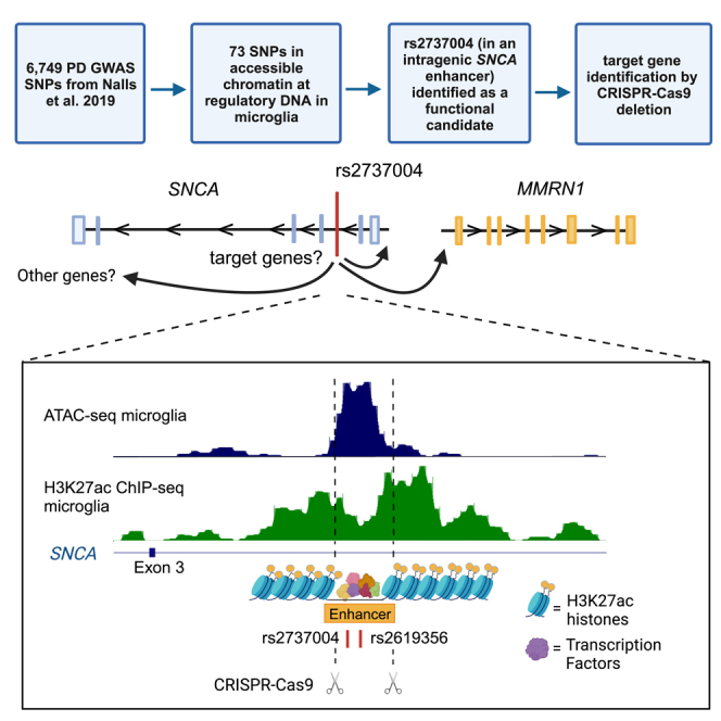
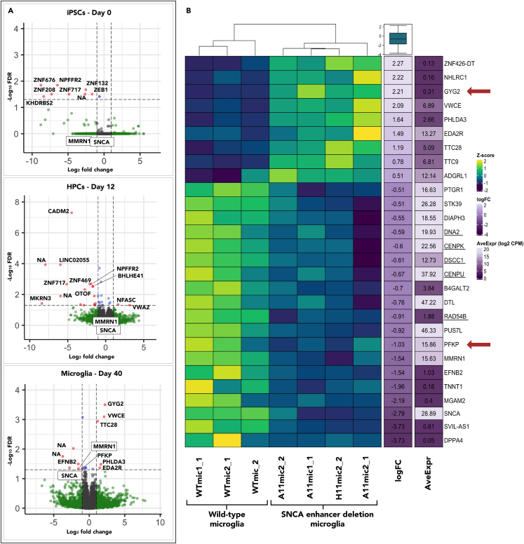
Genome-wide association studies have identified thousands of single nucleotide polymorphisms that associate with increased risk for Parkinson's disease (PD), but the functions of most of them are unknown. Using assay for transposase-accessible chromatin (ATAC) and H3K27ac chromatin immunoprecipitation (ChIP) sequencing data, we identified 73 regulatory elements in microglia that overlap PD risk SNPs. To determine the target genes of a "risk enhancer" within intron two of SNCA, we used CRISPR-Cas9 to delete the open chromatin region where two PD risk SNPs reside. The loss of the enhancer led to reduced expression of multiple genes including SNCA and the adjacent gene MMRN1. It also led to expression changes of genes involved in glucose metabolism, a process that is known to be altered in PD patients. Our work expands the role of SNCA in PD and provides a connection between PD-associated genetic variants and underlying biology that points to a risk mechanism in microglia.

Keywords: Biological sciences; Cellular neuroscience; Molecular neuroscience; Natural sciences; Neuroscience; Pathophysiology; Physiology.


Conflict of interest statement

The authors declare no competing interests.

Figures

None
Graphical abstract
Figure 1
Figure 1
A “risk haplotype” resides in an intragenic SNCA enhancer (A) Correlation analysis of the alleles of rs2737004 and rs2619356 using NCBI’s LDlink tools. The LDhap results show the frequency of each allele of the SNPs individually in all populations from NCBI. The colored boxes represent the haplotypes observed in the same population, with their counts and frequencies displayed below. The LDpair analysis reports the calculated statistics for linkage disequilibrium (D′ and R2) and the “goodness-of-fit” (chi-square and p value), which indicates the degree that the observed haplotype frequencies deviate from the expected allele frequencies. (B) Genome browser view of microglia ATAC- and H3K27ac ChIP-seq signals plotted with the locations of PD risk SNPs, all SNPs from 1,000 genomes, and the SNPs that are in LD with rs2737004. Rs2619356 was the only other SNP to overlap the ATAC-seq peak. Dotted lines represent the location of CRISPR/Cas9 guides designed to delete the 439 base pair region encompassing both SNPs in the “risk haplotype.”.
Figure 2
Figure 2
The SNCA risk enhancer shows evidence of functionality via 3D chromatin interactions and transcription factor binding (A) H3K27ac ChIP-seq tracks are displayed for microglia (purple) and neurons (green). Below are tracks showing PLAC-seq data for the same cell types. The red ovals denote primary interaction sites, in microglia, of the risk enhancer where rs2727004 is located. (B) MotifbreakR results showing transcription factor (TF) binding motifs of the TFs that have preference for the alleles of the risk haplotype (G for rs2737004 (left) and T for rs2619356 (right)). The letter size represents the results of the positional weight matrix that measures the frequency that the transcription factor binds to that nucleotide. In that same plot, the dashed black box demarcates the position of the SNP. The light blue boxes below represent the positions of the transcription factor binding motifs relative to the SNP’s genomic position, demarcated with the red box. (C) Remap ChIP-seq data for the transcription factors displayed in part B. The red lines show the position of each SNP within the ChIP-seq peak.
Figure 3
Figure 3
The SNCA risk enhancer controls the expression of SNCA, MMRN1, and a network of additional genes in microglia (A) EdgeR glmFit results comparing wild-type to SNCA-deletion cell lines across three time points in differentiation. Dotted lines represent a Log2 fold change of 2 and a false discover rate (FDR (Benjamini-Hochberg)) of 0.05. Gene name labels were manually added for consistency and clarity. See also Table S3 for results. (B). Heatmap of Z scores calculated from TMM-normalized log2 counts per million (log2 CPM). The genes in the heatmap represent those that have a Benjamini-Hochberg FDR cutoff of less than 0.1 (less stringent than used for the volcano plots in part A), and a fold change (FC) of over 1.4 (log FC > 0.5). The final plot was made by ranking the genes by log FC and taking the 15 genes at the top and bottom of that list.

References

    1. Nalls M.A., Blauwendraat C., Vallerga C.L., Heilbron K., Bandres-Ciga S., Chang D., Tan M., Kia D.A., Noyce A.J., Xue A., et al. Identification of novel risk loci, causal insights, and heritable risk for Parkinson's disease: a meta-analysis of genome-wide association studies. Lancet Neurol. 2019;18:1091–1102. - PMC - PubMed
    1. Gallagher M.D., Chen-Plotkin A.S. The Post-GWAS Era: From Association to Function. Am. J. Hum. Genet. 2018;102:717–730. - PMC - PubMed
    1. Soldner F., Stelzer Y., Shivalila C.S., Abraham B.J., Latourelle J.C., Barrasa M.I., Goldmann J., Myers R.H., Young R.A., Jaenisch R. Parkinson-associated risk variant in distal enhancer of alpha-synuclein modulates target gene expression. Nature. 2016;533:95–99. - PMC - PubMed
    1. Coetzee S.G., Pierce S., Brundin P., Brundin L., Hazelett D.J., Coetzee G.A. Enrichment of risk SNPs in regulatory regions implicate diverse tissues in Parkinson's disease etiology. Sci. Rep. 2016;6 - PMC - PubMed
    1. Pierce S.E., Booms A., Prahl J., van der Schans E.J.C., Tyson T., Coetzee G.A. Post-GWAS knowledge gap: the how, where, and when. NPJ Parkinsons Dis. 2020;6:23. - PMC - PubMed