Review
. 2024 Jan;29(Suppl 1):S11521.
doi: 10.1117/1.JBO.29.S1.S11521. Epub 2024 Feb 5.

Sounding out the dynamics: a concise review of high-speed photoacoustic microscopy

Affiliations
Review

Sounding out the dynamics: a concise review of high-speed photoacoustic microscopy

Soon-Woo Cho et al. J Biomed Opt. 2024 Jan.

Abstract

Significance: Photoacoustic microscopy (PAM) offers advantages in high-resolution and high-contrast imaging of biomedical chromophores. The speed of imaging is critical for leveraging these benefits in both preclinical and clinical settings. Ongoing technological innovations have substantially boosted PAM's imaging speed, enabling real-time monitoring of dynamic biological processes.

Aim: This concise review synthesizes historical context and current advancements in high-speed PAM, with an emphasis on developments enabled by ultrafast lasers, scanning mechanisms, and advanced imaging processing methods.

Approach: We examine cutting-edge innovations across multiple facets of PAM, including light sources, scanning and detection systems, and computational techniques and explore their representative applications in biomedical research.

Results: This work delineates the challenges that persist in achieving optimal high-speed PAM performance and forecasts its prospective impact on biomedical imaging.

Conclusions: Recognizing the current limitations, breaking through the drawbacks, and adopting the optimal combination of each technology will lead to the realization of ultimate high-speed PAM for both fundamental research and clinical translation.

Keywords: computational techniques; deep-learning; high-speed light source; high-speed scanning; optical acoustic detection; optoacoustic microscopy; photoacoustic microscopy.


Figures

Fig. 1
Fig. 1
Principle of PA imaging and penetration limits of PAM. (a) Illustration of PA imaging principle. (b) Penetration depth of OR-PAM and AR-PAM with different optical and acoustic focusing configurations. The images are reprinted with permission from Ref. .
Fig. 2
Fig. 2
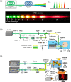
High-speed PAM using an SRS source. (a) Schematic illustration of the SRS effect in an optical fiber and the discrete Raman spectrum. (b) Schematic of the high-speed PAM system using an SRS source with an optical fiber pumped by a 1-MHz nanosecond pulsed laser. (c) Schematic of the high-speed PAM using an SRS source with a Raman crystal pumped by a picosecond pulsed laser. Images (a), (b), and (c) are reprinted with permission from Refs. , , and , respectively.
Fig. 3
Fig. 3
High-speed PAM system with mechanical scanning and optical scanning approaches. (a) Schematic of slider-crank scanner using two-channel PA probes. (b) High-resolution nude mouse ear PA image acquired by the slider-crank-scanner PAM system. (c) The illustration of high-speed PAM using a water-immersible polygon scanner with 12 facets. (d) PA images of the mouse brain vasculature and oxygenation over the entire cortex obtained from the polygon-scanner PAM system. Images (b) and (d) are reprinted from Refs.  and , respectively.
Fig. 4
Fig. 4
Various PA detection methods and their representative results by applying the optical scanning scheme. (a) Schematic of the planoconcave fiber-optic sensor. (b) in vivo PAM image of mouse ear obtained at 2 million A-lines per second. The scan area was 10  mm×10  mm, and the total acquisition time was 8 seconds. (c) Schematic of the fiber-laser based optic sensor method. (d) In vivo PAM image of the mouse ear at a C-scan frame rate of 0.2 Hz. The scanning FOV was 2.2×2.2  mm2. (e) Schematic of the transparent transducer method. (f) In vivo PAM image of whole cortical vasculature of the mouse brain. The total acquisition time was 5 s to scan an FOV of 6×6  mm2. Images (a), (b), and (c) are reproduced with permission from Refs. , , and , respectively.
Fig. 5
Fig. 5
Deep learning methods for upsampling PAM data. (a) PAM MAPs upsampled using FD U-Net with supervised training at various downsampling ratios (i.e., ratios [Dx, Dy]) compared with interpolation. (b) PAM MAPs upsampled using the unsupervised DIP iterative method compared with interpolation methods and FD U-Net. (c) PAM data upsampled in 3D using a combination of axial z-axis upsampling (SA-SRResNet) and lateral x-y-axis upsampling (DA-SRResNet). Images (a), (b), and (c) are reprinted with permission from Refs. , , and , respectively.
Fig. 6
Fig. 6
Improving PAM images by (a) aligning polygon facets in a fast polygon scanning system combined with deep learning FD U-Net upsampling, (b) recovering misaligned signal from bidirectional scan using deep learning, (c) correcting MEMS scanning distortion using a deep learning spatial weight matrix (SWM) with a dimensionality reduction, and (d) improving the image quality of low energy PAM using MT-RDN. Images (a) and (b)–(d) are reprinted with permission from Refs.  and –, respectively.

References

    1. Su J. L., et al. , “Advances in clinical and biomedical applications of photoacoustic imaging,” Expert Opin. Med. Diagn. 4(6), 497–510 (2010). 10.1517/17530059.2010.529127 - DOI - PMC - PubMed
    1. Xia J., Yao J., Wang L. V., “Photoacoustic tomography: principles and advances,” Electromagn. Waves 147, 1 (2014). 10.2528/PIER14032303 - DOI - PMC - PubMed
    1. Das D., et al. , “Another decade of photoacoustic imaging,” Phys. Med. Biol. 66(5), 05TR01 (2021). 10.1088/1361-6560/abd669 - DOI - PubMed
    1. Wang L. V., Yao J., “A practical guide to photoacoustic tomography in the life sciences,” Nat. Methods 13(8), 627–638 (2016). 10.1038/nmeth.3925 - DOI - PMC - PubMed
    1. Yao J., Wang L. V., “Photoacoustic microscopy,” Laser Photonics Rev. 7(5), 758–778 (2013). 10.1002/lpor.201200060 - DOI - PMC - PubMed

Publication types