. 2024 Feb 28;9(2):e0067823.
doi: 10.1128/msphere.00678-23. Epub 2024 Feb 7.

An obligate microsporidian parasite modulates defense against opportunistic bacterial infection in the yellow fever mosquito , Aedes aegypti

Affiliations

An obligate microsporidian parasite modulates defense against opportunistic bacterial infection in the yellow fever mosquito , Aedes aegypti

Noha K El-Dougdoug et al. mSphere. .

Abstract

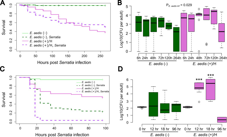
The ability of Aedes aegypti mosquitoes to transmit vertebrate pathogens depends on multiple factors, including the mosquitoes' life history traits, immune response, and microbiota (i.e., the microbes associated with the mosquito throughout its life). The microsporidium Edhazardia aedis is an obligate intracellular parasite that specifically infects Ae. aegypti mosquitoes and severely affects mosquito survival and other life history traits critical for pathogen transmission. In this work, we investigated how E. aedis impacts bacterial infection with Serratia marcescens in Ae. aegypti mosquitoes. We measured development, survival, and bacterial load in both larval and adult stages of mosquitoes. In larvae, E. aedis exposure was either horizontal or vertical and S. marcescens was introduced orally. Regardless of the route of transmission, E. aedis exposure resulted in significantly higher S. marcescens loads in larvae. E. aedis exposure also significantly reduced larval survival but subsequent exposure to S. marcescens had no effect. In adult females, E. aedis exposure was only horizontal and S. marcescens was introduced orally or via intrathoracic injection. In both cases, E. aedis infection significantly increased S. marcescens bacterial loads in adult female mosquitoes. In addition, females infected with E. aedis and subsequently injected with S. marcescens suffered 100% mortality which corresponded with a rapid increase in bacterial load. These findings suggest that exposure to E. aedis can influence the establishment and/or replication of other microbes in the mosquito. This has implications for understanding the ecology of mosquito immune defense and potentially disease transmission by mosquito vector species.

Importance: The microsporidium Edhazardia aedis is a parasite of the yellow fever mosquito, Aedes aegypti. This mosquito transmits multiple viruses to humans in the United States and around the world, including dengue, yellow fever, and Zika viruses. Hundreds of millions of people worldwide will become infected with one of these viruses each year. E. aedis infection significantly reduces the lifespan of Ae. aegypti and is therefore a promising novel biocontrol agent. Here, we show that when the mosquito is infected with this parasite, it is also significantly more susceptible to infection by an opportunistic bacterial pathogen, Serratia marcescens. This novel discovery suggests the mosquito's ability to control infection by other microbes is impacted by the presence of the parasite.

Keywords: Aedes aegypti; Edhazardia aedis; Serratia marcescens; horizontal transmission; immune defense; immune response; immunity; microsporidia; parasite; vertical transmission.


Conflict of interest statement

The authors declare no conflict of interest.

Figures

Fig 1
Fig 1
Life cycle of the microsporidia Edhazardia aedis in Ae. aegypti mosquitoes. In horizontal transmission, infective uninucleate spores of E. aedis are ingested by Ae. aegypti larvae from the aquatic environment. These spores invade the larval midgut lumen and develop into binucleate spores. Infected adults eclose and binucleate spores of E. aedis infect oenocytes of adult females. They are then transmitted through the hemocoel to the ovaries where they are vertically transmitted to offspring. When larvae hatch from infected eggs, they experience a highly virulent infection by E. aedis primarily in the fat body, which leads to the death of larvae and the release of infective uninucleate spores in the aquatic environment. These spores are ingested by susceptible larvae to complete the cycle. This image was created with BioRender (https://www.biorender.com/).
Fig 2
Fig 2
Larvae infected vertically with E. aedis have significantly higher numbers of Serratia bacteria. Ae. aegypti larvae were either uninfected [E. aedis(−)] or vertically infected with E. aedis via the infected mother [E. aedis(+)/V)] and then exposed orally to S. marcescens-GFP early in the 4th instar (Fig. S1). Oral exposure lasted for 6 hours at which point larvae were washed and transferred to clean water. Data were collected from a total of two replicate experiments. In each replicate, starting sample sizes were n = 50 mosquitoes per treatment group. (A) Pupation was significantly affected by E. aedis (P = 6.41 × 10⁻⁶) but not Serratia (P = 0.241) infection, and there was no significant interaction between these predictor variables (P = 0.8969). (B) Eclosion was significantly affected by E. aedis (P = 2.15 × 10⁻¹⁰) but not Serratia (P = 0.9033) infection, and there was no significant interaction between these predictor variables (P = 0.5976). (C) Overall survival was significantly affected by E. aedis (P < 2 × 10⁻¹⁶) but not Serratia (P = 0.60) infection. We were unable to test for an interaction, but the survival curves suggest no interaction given the highly similar effects of Serratia regardless of E. aedis infection status. (D) Serratia bacterial load was significantly higher in E. aedis(+) mosquitoes (P = 1.135 × 10⁻⁷), and this was consistent at all time points post-Serratia exposure (E. aedis × hour interaction, P = 0.160). Each box plot represents n = 10 mosquitoes, and the P-value indicates the overall effect of E. aedis infection on bacterial load.
Fig 3
Fig 3
Larvae infected horizontally with E. aedis have significantly higher numbers of Serratia bacteria. Ae. aegypti larvae were either uninfected [E. aedis(−)] or horizontally exposed to E. aedis spores in larval water [E. aedis(+)/H] and then exposed orally to S. marcescens-GFP (Fig. S2). Oral exposure lasted for 6 hours at which point larvae were washed and transferred to clean water. Data were collected from a total of two replicate experiments. In each replicate, starting sample sizes were n = 50 mosquitoes per treatment group. (A) Pupation was significantly slowed by E. aedis (P = 3.39 × 10⁻⁴) but was not affected by Serratia (P = 0.8669) infection, and there is a marginally significant interaction between E. aedis and Serratia infection on pupation rate (P = 0.0484). (B) Eclosion was significantly affected by E. aedis (P = 1.07 × 10⁻³) but not Serratia (P = 0.6391) infection, and there was no significant interaction between these predictor variables (P = 0.5004). (C) Overall survival was significantly affected by E. aedis (P = 1.00 × 10⁻¹¹) but not Serratia (P = 0.9) infection. We were unable to test for an interaction, but the survival curves suggest no interaction given the highly similar effects of Serratia regardless of E. aedis infection status. (D) Serratia load was significantly higher in E. aedis(+)/H individuals compared to E. aedis(−) individuals, but this varied over time (E. aedis × hour interaction, P = 0.015). Serratia load was higher at 48 hours (P = 0.003) and 72 hours (P = 7.06 × 10⁻⁴) but not at 6, 24, or 96 hours post-initial Serratia exposure. Each box plot represents n = 10 mosquitoes, and asterisks denote significant differences from the E. aedis(−) treatment at the corresponding time point.
Fig 4
Fig 4
Infection of Serratia through intrathoracic injection has a significant effect on the mortality rate and bacterial load of adult females infected horizontally with E. aedis. Adult females infected horizontally with E. aedis [E. aedis (+)/H] and uninfected females [E. aedis(−)] were infected with S. marcescens-GFP either orally in a sugar meal (oral exposure lasted for 6 hours) (Fig. S3) or through intrathoracic injection (Fig. S4). Data were collected from a total of two replicate experiments. In each replicate, starting sample sizes were n = 50 mosquitoes per treatment group. Under Serratia oral infection, (A) overall survival was significantly affected by E. aedis (P = 2 × 10⁻¹⁶) and there was no effect of Serratia on survival in either E. aedis(+) (P = 0.64) or the E. aedis(−) groups (P = 0.98). (B) Serratia bacterial load was significantly higher in E. aedis(+) mosquitoes (P = 0.029), and this was consistent at all time points post-Serratia exposure (E. aedis × hour interaction, P = 0.680). Each box plot represents n = 10 mosquitoes, and the P-value indicates the overall effect of E. aedis treatment on bacterial load. Under Serratia infection through intrathoracic injection, (C) E. aedis (P = 4.0 × 10⁻¹³) and Serratia (P = 2.0 × 10⁻¹⁶) both significantly reduced survival. The effect of Serratia was significant in both E. aedis(+) (P = 3.1 × 10⁻¹⁴) and the E. aedis(−) groups (P < 2.0 × 10⁻¹⁶) considered separately. The mortality rate was highest in individuals co-infected with E. aedis and Serratia (100% mortality by 24 hours). (D) E. aedis significantly affected Serratia load (P = 8.14 × 10⁻⁸) but this effect varied over time (E. aedis × hour interaction, P = 2.29 × 10⁻⁵). E. aedis(+) females had significantly higher bacterial loads at 12 hours (P = 1.69 × 10⁻³) and 18 hours (P = 4.75 × 10⁻⁴) post-infection. Sample sizes were n = 10 for all time points except: E. aedis(−)/96 hours, n = 8; E. aedis(+)/18 hours, n = 6; E. aedis(+)/96 hours, n = 2. Asterisks denote significant differences from the E. aedis(−) treatment at the corresponding time point.

References

    1. Centers for disease control and prevention, “mosquito-borne diseases". 2016. www.cdc.gov/niosh/topics/outdoor/mosquito-borne/default.html.
    1. Powell JR. 2018. Mosquito-borne human viral diseases: why Aedes aegypti. Am J Trop Med Hyg 98:1563–1565. doi:10.4269/ajtmh.17-0866 - DOI - PMC - PubMed
    1. Brady OJ, Hay SI. 2020. The global expansion of dengue: how Aedes aegypti mosquitoes enabled the first pandemic arbovirus. Annu Rev Entomol 65:191–208. doi:10.1146/annurev-ento-011019-024918 - DOI - PubMed
    1. Gubler DJ. 2002. The global emergence/resurgence of arboviral diseases as public health problems. Arch Med Res 33:330–342. doi:10.1016/s0188-4409(02)00378-8 - DOI - PubMed
    1. World health organization . 2023. Dengue and severe dengue. Available from: https://www.who.int/news-room/fact-sheets/detail/dengue-and-severe-dengue

Supplementary concepts