. 2024 Jun;23(3):911-925.
doi: 10.1007/s10237-023-01813-3. Epub 2024 Feb 7.

Uniaxial mechanical stretch properties correlated with three-dimensional microstructure of human dermal skin

Affiliations

Uniaxial mechanical stretch properties correlated with three-dimensional microstructure of human dermal skin

Mengyao Zhou et al. Biomech Model Mechanobiol. 2024 Jun.

Abstract

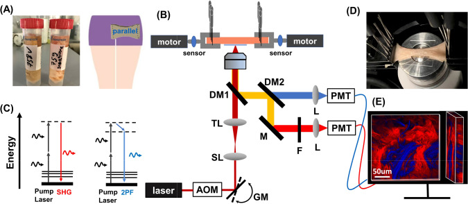
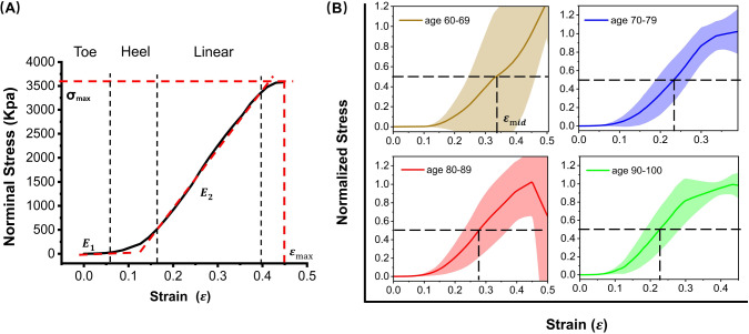
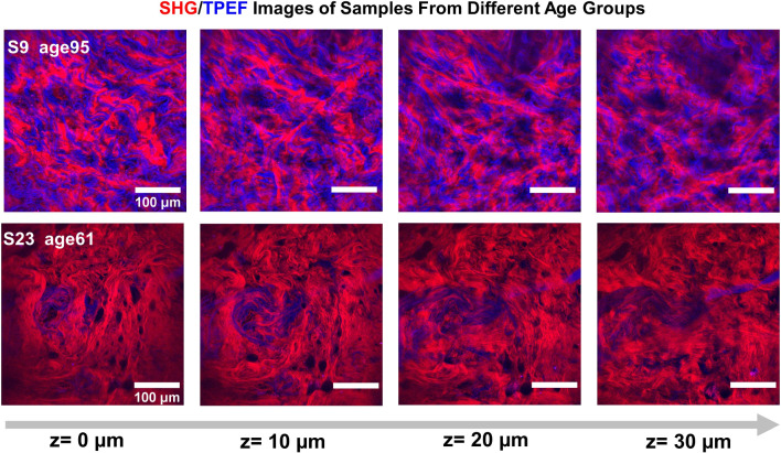
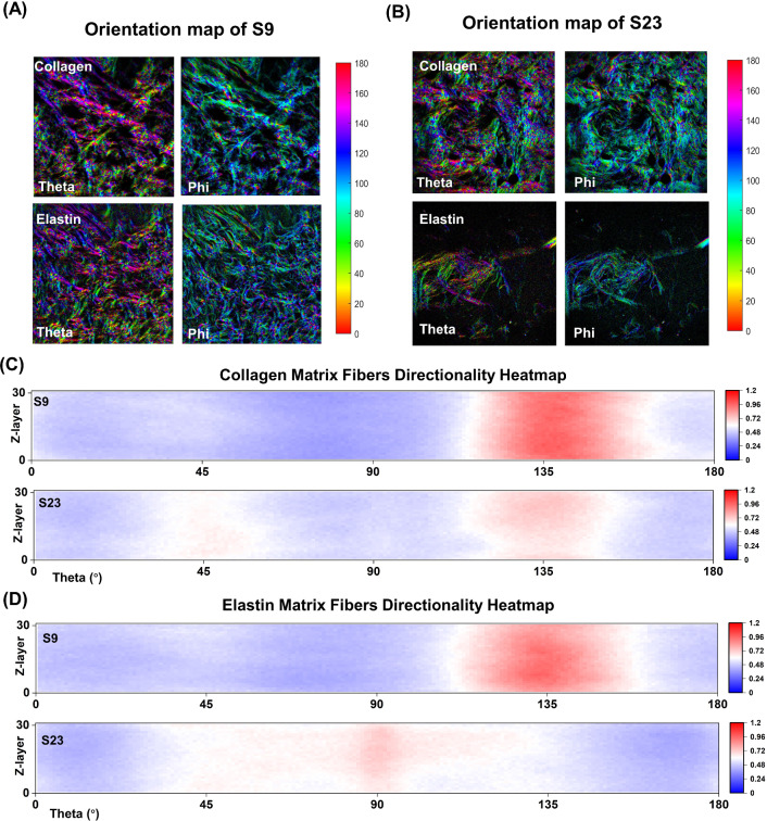
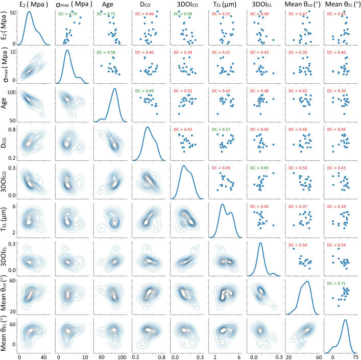
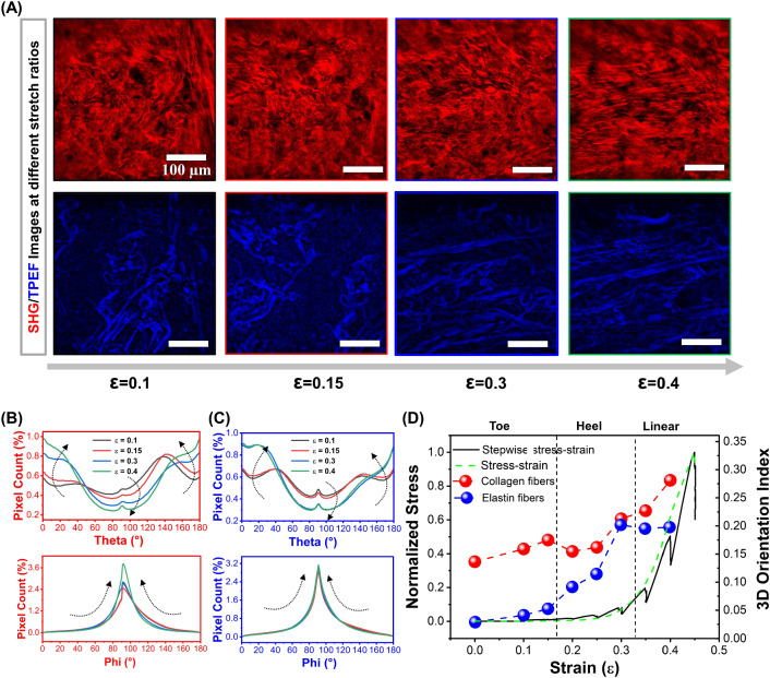
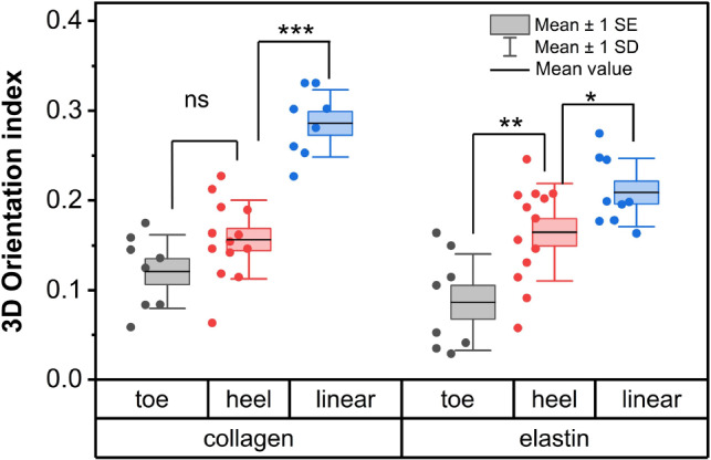
The intact and healthy skin forms a barrier to the outside world and protects the body from mechanical impact. The skin is a complex structure with unique mechano-elastic properties. To better direct the design of biomimetic materials and induce skin regeneration in wounds with optimal outcome, more insight is required in how the mechano-elastic properties emerge from the skin's main constituents, collagen and elastin fibers. Here, we employed two-photon excited autofluorescence and second harmonic generation microscopy to characterize collagen and elastin fibers in 3D in 24 human dermis skin samples. Through uniaxial stretching experiments, we derive uni-directional mechanical properties from resultant stress-strain curves, including the initial Young's modulus, elastic Young's modulus, maximal stress, and maximal and mid-strain values. The stress-strain curves show a large variation, with an average Young's modules in the toe and linear regions of 0.1 MPa and 21 MPa. We performed a comprehensive analysis of the correlation between the key mechanical properties with age and with microstructural parameters, e.g., fiber density, thickness, and orientation. Age was found to correlate negatively with Young's modulus and collagen density. Moreover, real-time monitoring during uniaxial stretching allowed us to observe changes in collagen and elastin alignment. Elastin fibers aligned significantly in both the heel and linear regions, and the collagen bundles engaged and oriented mainly in the linear region. This research advances our understanding of skin biomechanics and yields input for future first principles full modeling of skin tissue.

Keywords: Collagen fibers; Elastin fibers; Human skin; Mechanical properties; Second harmonic generation; Uniaxial skin stretch.


Conflict of interest statement

The authors declare that there are no conflicts of interest related to this article.

Figures

Fig. 1
Fig. 1
A Human skin samples from cadaver thighs and cutting orientation. B Schematic diagram of the experimental setup: The laser has a central wavelength of 1050 nm with pulse duration less than 80 fs; AOM acousto-optic modulator, GM galvo mirrors; SL scan lens, TL tube lens with adjustable focus, DM1 dichroic mirror reflecting backscattering signals from fundamental photons, DM2 dichroic mirror splitting TPEF and SHG channels, M mirror, F bandpass filter, F520/35, L focusing lens, PMT photomultiplier tube detectors. C Energy level diagram of the SHG and TPEF process. D Image shows the deformation of skin sample at 115% stretching. E 3D volume image of collagen and elastin fibers from human thigh skin. Red color represents collagen fibers and blue color represents elastin fibers
Fig. 2
Fig. 2
Uniaxial stretching data for human thigh dermis skin. A A representative stress–strain curve of human dermis skin (S5), where E1 is the initial Young’s modulus, the slope of the curve in the toe region, E2 is the elastic modulus (Young’s modulus), the slope of the linear region of the curve, and σmax and εmax are maximum stress and maximum strain before failure. B Average normalized stress–strain curves for different age groups of donors, with shaded areas representing standard deviations. The dashed lines illustrate 50% of the maximum stress, corresponding to the strain denoted as εmid. The individual stress–strain curves of each group can be found in SI (Figure S1)
Fig. 3
Fig. 3
Pre-stretching images of collagen and elastin fibers of sample 9 and 23 at different depths. Collagen fibers in red color, elastin fibers in blue color. FOV: 400 × 400 μm2. The presented images were processed with CLAHE. The intensity histograms of raw images and processed images of S9 and S23 at different imaging depths can be found in SI of Figure S2 and S3
Fig. 4
Fig. 4
The orientation map of the collagen and elastin fibers of A sample 9, B sample 23 in the middle layer of the whole z-stack. Series of heatmaps represent the directional orientation of C collagen and D elastin fibers oriented between 1 and 180 degrees across the z-layer for 2 samples (S9, S23). The heatmap represents the orientation of collagen and elastin fiber as percentage of pixel count. As indicated by the scale bar on the right of each heatmap, the redder the color, the higher the counts. The z-layers are ranged from bottom to top in ascending order
Fig. 5
Fig. 5
The upper half of the diagram shows the scatter plots between the various variables and the distance correlation in green (p<0.05) and red (p>0.05). On the diagonal the kernel density estimates of the single variable distributions are shown. The lower half of the diagram shows the isolines of the kernel density estimates of the various bivariate distributions. The distributions and correlations between all variables can be found in SI (Figure S3).E2: Elastic Young’s modulus, σmax maximum stress, age of the donor; DCO, density of collagen fibers; 3DOICO, collagen 3D orientation index; TEL, elastin fiber thickness; 3DOIEL, elastin 3D orientation index; main θCO, main θ angle of collagen fibers, main θEL, and main θ angle of elastin fibers
Fig. 6
Fig. 6
Evolution of collagen and elastin fiber orientation of a human thigh dermis skin sample during stretching (sample information: female, age 88, S22). A Representative 2D images of collagen fibers (first row) and elastin fibers (second row) at different strain, which are extracted from corresponding 3D image stacks. FOV: 400×400μm2 (clip off dark invalid areas around the edges from a 500×500μm2 raw data). 3D image stacks at different strain can be seen in the SI (visualization 1, 2, 3 and 4 for ε = 0.1, 0.15, 0.3 and 0.4, respectively), and a time-lapse video monitoring the changes of the fibers during stretching can be seen in visualization 5. θ and φ angular distribution of B collagen and C elastin fibers across the z-stack at different strain. D The 3DOI of collagen and elastic fibers with increasing strain, and the corresponding stress–strain curves, with solid black line indicating the stress–strain curve with stops for imaging, and the dashed green line for the stress–strain curve measured prior to imaging at a continuous stretch speed of 0.5 mm/s. The dashed black lines indicate the toe, heel, and linear regions (Fratzl et al. ; Gutsmann et al. 2004)
Fig. 7
Fig. 7
3DOI of collagen and elastin fibers with dynamic stress–strain curves. First row is low-strain midpoint group, A S11, B S13, C S14. Second row is high-strain midpoint group D S21, E S22, F S24
Fig. 8
Fig. 8
The box plot of mean value of 3DOI at different stress regions of collagen and elastin fibers. * stands for p<0.05, ** for p<0.01, *** for p<0.001 and ns for no significant difference

References

    1. Allain Jean-Marc, Lynch Barbara, Schanne-Klein Marie-Claire (2019) Multiscale characterisation of skin mechanics through in situ imaging. Skin Biophys From Exp Charact Adv Modell pp. 235–263
    1. Bancelin Stéphane, Lynch Barbara, Bonod-Bidaud Christelle, Ducourthial Guillaume, Psilodimitrakopoulos Sotiris, Dokládal Petr, Allain Jean-Marc, Schanne-Klein Marie-Claire, Ruggiero Florence. Ex vivo multiscale quantitation of skin biomechanics in wild-type and genetically-modified mice using multiphoton microscopy. Sci Rep. 2015;5(1):17635. doi: 10.1038/srep17635. - DOI - PMC - PubMed
    1. Blair Michael J, Jones Jake D, Woessner Alan E, Quinn Kyle P. Skin structure-function relationships and the wound healing response to intrinsic aging. Adv Wound Care. 2020;9(3):127–143. doi: 10.1089/wound.2019.1021. - DOI - PMC - PubMed
    1. Chow Ming-Jay, Turcotte Raphaël, Lin Charles P, Zhang Yanhang. Arterial extracellular matrix: a mechanobiological study of the contributions and interactions of elastin and collagen. Biophys J. 2014;106(12):2684–2692. doi: 10.1016/j.bpj.2014.05.014. - DOI - PMC - PubMed
    1. Daly Colin H. Biomechanical properties of dermis. J Investig Dermatol. 1982;79(1):17–20. doi: 10.1038/jid.1982.4. - DOI - PubMed

LinkOut - more resources