

Long-term clinical outcomes of patients with sympathetic ophthalmia
- PMID: 38324101
- PMCID: PMC10850007
- DOI: 10.1007/s10792-024-03007-x
Long-term clinical outcomes of patients with sympathetic ophthalmia
Abstract
Purpose: To present the long-term clinical outcomes of patients with sympathetic ophthalmia (SO).
Methods: Retrospective review of patients' medical files between 2002 and 2022.
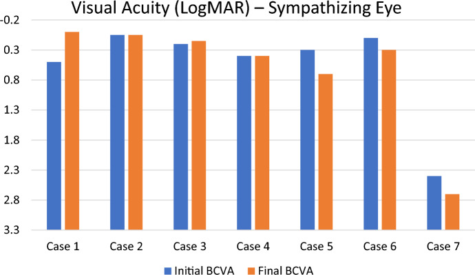
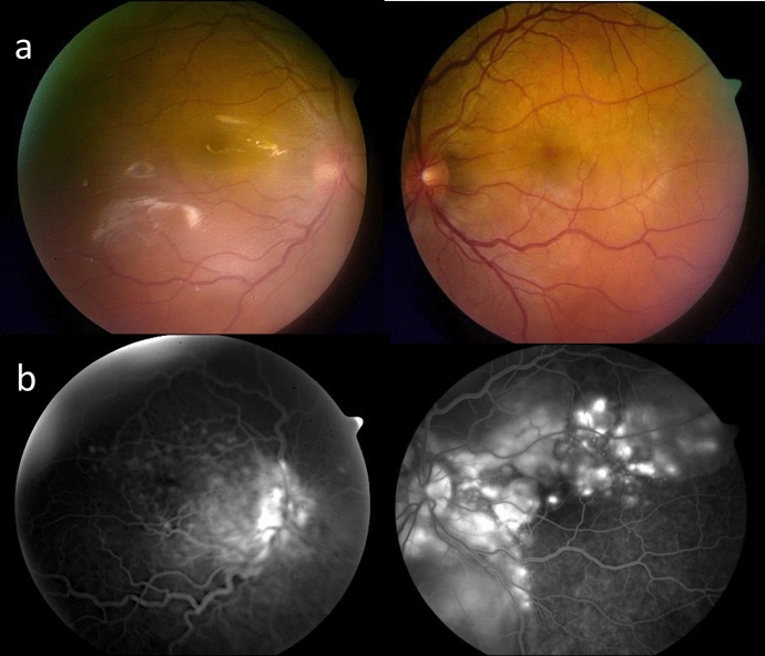
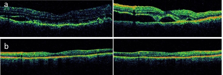
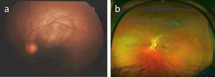
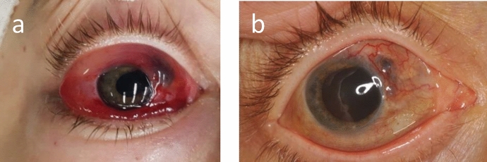
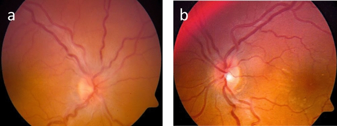
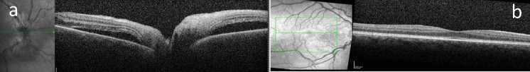
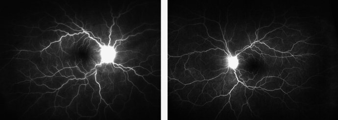
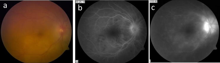
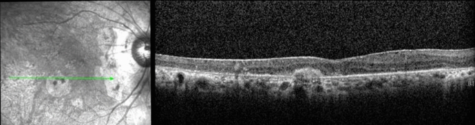
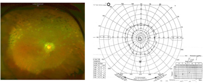
Results: Included were seven patients (four males). The mean ± SD age at presentation was 37.9 ± 22.5 years. Four patients had co-morbidities: three had diabetes mellitus type 2 and one had Turner Syndrome. Trauma was the inciting event in six patients and postoperative endophthalmitis in one patient. Decreased visual acuity (VA) was the leading symptom in the sympathizing eye and all of the patients presented with panuveitis. The mean ± SD interval between the triggering incident and the onset of SO in six cases was 4.3 ± 4.2 months. One case presented 30 years following the eye injury. Five patients underwent enucleation/evisceration of the exciting eye. The mean ± SD presenting LogMAR BCVA in the sympathizing eye was 0.57 ± 0.82, and the final LogMAR BCVA was 0.61 ± 0.95. Inflammation was completely controlled in 5 patients at a mean ± SD of 8.55 ± 9.21 months following the institution of immunomodulatory therapy, and it was partially controlled in 2 patients. VA deteriorated in all 3 diabetic patients and improved or remained stable in the 4 young and healthy patients. The mean ± SD follow-up period after achieving drug-free remission was 28 ± 22.8 months. The mean ± SD follow-up time was 6.8 ± 5.6 years.
Conclusions: SO is one of the most sight-threatening conditions, affecting the healthy eye. In this cohort, the favorable visual outcome was especially seen in young and healthy individuals. Visual prognosis is directly related to prompt diagnosis and treatment.
Keywords: Dalen-Fuchs nodules; Granulomatous uveitis; Ocular trauma; Sympathetic ophthalmia.
© 2024. The Author(s).
Conflict of interest statement
The authors do not have any financial or non-financial interests that are relevant to disclose.
Figures
References
-
- He B, Tanya SM, Wang C, Kezouh A, Torun N, Ing E (2022) The incidence of sympathetic ophthalmia after trauma: a meta-analysis. Am J Ophthalmol 234:117–125. 10.1016/j.ajo.2021.06.036 - PubMed
-
- Muccioli C, Oden N, Okada AA, Palestine AG, Rao NA, Read RW, Thorne JE, Trusko BE (2021) Classification criteria for sympathetic ophthalmia. Am J Ophthalmol. 10.1016/j.ajo.2021.03.048
-
- Patel SS, Dodds EM, Echandi LV, Couto CA, Schlaen A, Tessler HH, Goldstein DA (2014) Long-term, drug-free remission of sympathetic ophthalmia with high-dose, short-term chlorambucil therapy. Ophthalmology 121(2):596–602. 10.1016/j.ophtha.2013.09.009 - PubMed
-
- Lubin JR, Albert DM, Weinstein M (1980) Sixty-five years of sympathetic ophthalmia: a clinicopathologic review of 105, cases (1913–1978). Ophthalmology 87(2):109–121. 10.1016/S0161-6420(80)35270-6 - PubMed
MeSH terms
LinkOut - more resources
Full Text Sources
Medical
