. 2024 Feb 7;44(2):60.
doi: 10.1007/s10875-024-01660-6.

Interface Gain-of-Function Mutations in TLR7 Cause Systemic and Neuro-inflammatory Disease

Affiliations

Interface Gain-of-Function Mutations in TLR7 Cause Systemic and Neuro-inflammatory Disease

Clémence David et al. J Clin Immunol. .

Abstract

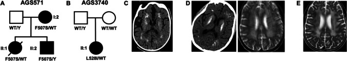
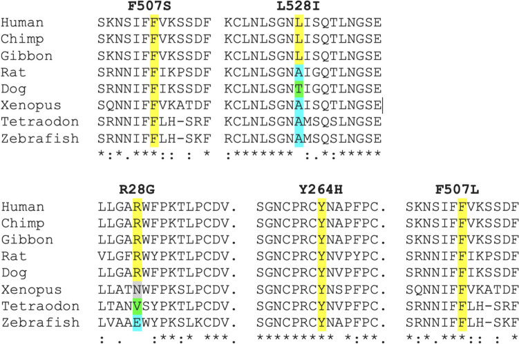
TLR7 recognizes pathogen-derived single-stranded RNA (ssRNA), a function integral to the innate immune response to viral infection. Notably, TLR7 can also recognize self-derived ssRNA, with gain-of-function mutations in human TLR7 recently identified to cause both early-onset systemic lupus erythematosus (SLE) and neuromyelitis optica. Here, we describe two novel mutations in TLR7, F507S and L528I. While the L528I substitution arose de novo, the F507S mutation was present in three individuals from the same family, including a severely affected male, notably given that the TLR7 gene is situated on the X chromosome and that all other cases so far described have been female. The observation of mutations at residues 507 and 528 of TLR7 indicates the importance of the TLR7 dimerization interface in maintaining immune homeostasis, where we predict that altered homo-dimerization enhances TLR7 signaling. Finally, while mutations in TLR7 can result in SLE-like disease, our data suggest a broader phenotypic spectrum associated with TLR7 gain-of-function, including significant neurological involvement.

Keywords: TLR7; stem cell transplantation; systemic lupus erythematosus.


Conflict of interest statement

Y.J.C. is an associate editor of the Journal of Clinical Immunology. The authors declare no other conflicts of interest, financial or otherwise.

Figures

Fig. 1
Fig. 1
Genetic and clinical data. Family pedigrees (A AGS571; B AGS3740) where an affected individual harbors a rare non-synonymous missense substitution in TLR7, in either the hemizygous (male) or heterozygous (female) state. Circles and squares indicate female and male family members, respectively. Filled shapes indicate affected status. The line across II:1 in AGS571 indicates deceased status. WT = wild-type. C Cranial CT in AGS571 II:2 at the age of < 1 year. D Cranial CT (left) and axial T2-weighted MRI (right) of AGS3740 II:1 at age 10 years. E Progression of high signal in the deep white matter of AGS3740 II:1 by age 11 years (1 year after hematopoietic stem cell transplantation)
Fig. 2
Fig. 2
Clustal Omega alignment of TLR7. Clustal Omega alignment of TLR7 with identified non-synonymous missense substitutions highlighted. Top: substitutions identified in this study. Bottom: substitutions identified in Brown et al. Alignments are based on the human transcript of TLR7: ENST00000380659.4/NM_016562.4; NP_057646.1
Fig. 3
Fig. 3
In vitro assessment of TLR7 signaling. NF-κB luciferase reporter activity following co-transfection of HEK293T cells with empty vector (EV), wild-type (WT) TLR7 or variants of TLR7, and UNC93B1 plasmid and stimulation with R848 0.1 µg/mL. Data are normalized to WT response to R848. Mean ± SEM of n = 3 experiments. Two-way ANOVA with Dunnett’s post hoc test: **p < 0.01, *p < 0.5. NS: non-stimulated
Fig. 4
Fig. 4
Modeling of TLR7 and assessment of identified variants. A Top view and side views of TLR7 structure based upon PDB ID 7cyn, with the location of indicated amino acids (variants at F507 and L528 found in this study) highlighted. B Predicted change in the Gibbs free energy of folding (ΔΔG) of TLR7 variants measured in monomeric TLR7 structures (left). Predicted intermolecular ΔΔG defined as the difference between the ΔΔG of the TLR7 monomer and the dimer (right). Box, 25th and 75th percentiles; middle line, median; whiskers, 1.5 times the interquartile range. C Violin plot showing the average buried surface areas of residues at which gnomAD (v4) variants are recorded in TLR7 versus the 3 residues of the 4 variants identified in this study or by Brown et al. [10]. P-value was calculated with a two-sided Wilcoxon rank-sum test (right)

References

    1. Dorner T, Furie R. Novel paradigms in systemic lupus erythematosus. Lancet. 2019;393(10188):2344–2358. doi: 10.1016/S0140-6736(19)30546-X. - DOI - PubMed
    1. Omarjee O, Picard C, Frachette C, Moreews M, Rieux-Laucat F, Soulas-Sprauel P, et al. Monogenic lupus: dissecting heterogeneity. Autoimmun Rev. 2019;18(10):102361. doi: 10.1016/j.autrev.2019.102361. - DOI - PubMed
    1. Vinuesa CG, Shen N, Ware T. Genetics of SLE: mechanistic insights from monogenic disease and disease-associated variants. Nat Rev Nephrol. 2023;19(9):558–572. doi: 10.1038/s41581-023-00732-x. - DOI - PubMed
    1. Zhang SY, Jouanguy E, Ugolini S, Smahi A, Elain G, Romero P, et al. TLR3 deficiency in patients with herpes simplex encephalitis. Science. 2007;317(5844):1522–1527. doi: 10.1126/science.1139522. - DOI - PubMed
    1. Zhang Q, Bastard P, Liu Z, Le Pen J, Moncada-Velez M, Chen J, et al. Inborn errors of type I IFN immunity in patients with life-threatening COVID-19. Science. 2020;370(6515):eabd4570. - PMC - PubMed

Publication types