Case Reports
. 2024 Aug;23(4):1498-1508.
doi: 10.1007/s12311-024-01666-1. Epub 2024 Feb 7.

Comprehensive Analysis of a Japanese Pedigree with Biallelic ACAGG Expansions in RFC1 Manifesting Motor Neuronopathy with Painful Muscle Cramps

Affiliations
Case Reports

Comprehensive Analysis of a Japanese Pedigree with Biallelic ACAGG Expansions in RFC1 Manifesting Motor Neuronopathy with Painful Muscle Cramps

Rumiko Izumi et al. Cerebellum. 2024 Aug.

Abstract

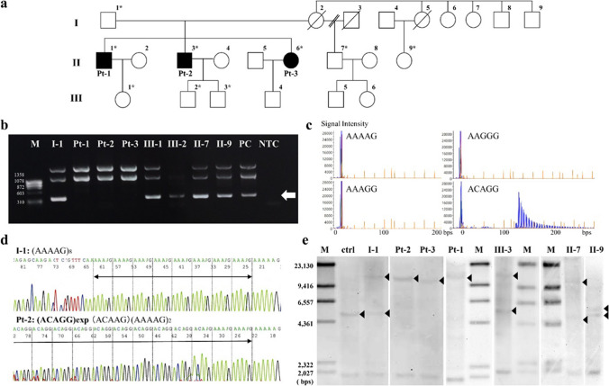
Cerebellar ataxia, neuropathy, and vestibular areflexia syndrome (CANVAS) is an autosomal recessive multisystem neurologic disorder caused by biallelic intronic repeats in RFC1. Although the phenotype of CANVAS has been expanding via diagnostic case accumulation, there are scant pedigree analyses to reveal disease penetrance, intergenerational fluctuations in repeat length, or clinical phenomena (including heterozygous carriers). We identified biallelic RFC1 ACAGG expansions of 1000 ~ repeats in three affected siblings having sensorimotor neuronopathy with spinocerebellar atrophy initially presenting with painful muscle cramps and paroxysmal dry cough. They exhibit almost homogeneous clinical and histopathological features, indicating motor neuronopathy. Over 10 years of follow-up, painful intractable muscle cramps ascended from legs to trunks and hands, followed by amyotrophy and subsequent leg pyramidal signs. The disease course combined with the electrophysical and imagery data suggest initial and prolonged hyperexcitability and the ensuing spinal motor neuron loss, which may progress from the lumbar to the rostral anterior horns and later expand to the corticospinal tract. Genetically, heterozygous ACAGG expansions of similar length were transmitted in unaffected family members of three successive generations, and some of them experienced muscle cramps. Leukocyte telomere length assays revealed comparatively shorter telomeres in affected individuals. This comprehensive pedigree analysis demonstrated a non-anticipating ACAGG transmission and high penetrance of manifestations with a biallelic state, especially motor neuronopathy in which muscle cramps serve as a prodromal and disease progress marker. CANVAS and RFC1 spectrum disorder should be considered when diagnosing lower dominant motor neuron disease, idiopathic muscle cramps, or neuromuscular hyperexcitability syndromes.

Keywords: RFC1; ACAGG; CANVAS; Motor neuronopathy; Muscle cramp; Telomere.


Conflict of interest statement

The authors have no conflicts of interest to declare.

Figures

Fig. 1
Fig. 1
Family pedigree and genetic analysis. The family pedigree is shown (a). The family includes three affected individuals with nearly identical manifestations (II-1, II-3, II-6, corresponding to Pt-1, Pt-2, and Pt-3, respectively). Their mother (I-2) complained of muscle cramps and gait disturbances but with insufficient medical information. Non-symptomatic heterozygous carriers of I-1 and II-7 underwent comprehensive examinations that did not show any neurological deficit. Isolated muscle cramps had been recognized in III-1–4 from their childhood to their 30 s. These muscle cramps were essentially non-progressive, and the neurological examination of III-2 and III-3 revealed no sign of the disease. Asterisks indicate individuals whose DNA was used for genetic analyses. The RFC1 intron 2 short-range flanking PCR demonstrated absent amplification in Pt-1–3, which is estimated to be 348 bps in length (b, indicated by an arrow). Per the repeat-primed PCR that was performed, the ladder pattern is only detected via ACAGG-tagged primers in I-1, Pt-1–3, II-7, III-1, and III-2, except in II-9 (c). From Sanger-sequencing following nested PCR, I-1 has a referential allele (d, shown above), whereas Pt-2 has homozygous expansion of ACAGG motif (d, shown below). Per Southern blot analyses (e), a wild-type allele fragment supposed to be 5037 bp long is absent in Pt-1–3, and a homozygous single band approximately ranging from 10 to 15 kbps can be seen alternatively. Both expanded and wild-type bands are seen in I-1, II-7, and III-3
Fig. 2
Fig. 2
Muscle computed tomography. Muscle computed tomography was evaluated twice in Pt-II at the ages of 57 and 71. The images show slices of the forearm and arm (a, left and right), neck (b), thorax (c), abdomen (d), pelvis (e), thigh (f), and distal legs (g). Symmetrical fatty changes in the medial head and to a lesser extent the lateral head of the gastrocnemius are present at 57 years (g, left panel), whereas pathological muscle atrophy cannot be identified in other levels. At 71 years, all lower leg muscles were severely fatty-replaced, relatively sparing the tibialis anterior and extensor digitorum longus muscles (g, right panel). The thigh exhibited moderate and diffuse atrophy; however, the hamstring muscles are predominantly affected (f, right panel). In the truncus, paraspinal muscles are fatty-degenerated continuously from the cervical level to the lumbar level. In the upper extremities, diffuse muscle atrophy is more prominent in the forearms than in the arms (a, right panel)
Fig. 3
Fig. 3
Electrophysiology. F-waves induced by left median nerve stimulation in Pt-2 at the age of 71 are shown (a). Whereas the amplitude of the CMAP and motor conduction is maintained at the normal level, the occurrence of F-waves is reduced to 31% occupied by repeater F-waves. The F/M amplitude ratio is increased to 0.4. The nEMG performed on the left gastrocnemius in Pt-3 is shown (b). Motor units of simple forms as large as 14 mV are recruited by voluntary contraction. The polysomnography of Pt-3 reveals frequent periodic limb movements (PLMs) that are monitored via both tibialis anteriors (TAs), which frequently occurred early during sleep (c) and occurred a total of 219 times during non-rapid-eye-movement sleep. The apnea–hypopnea index, lowest SpO2, and maximum apnea duration were 6.7/h, 82%, and 44 s, respectively, indicating mild sleep apnea. RIP: respiratory inductance plethysmography
Fig. 4
Fig. 4
Pathology of muscle biopsy. Hematoxylin–eosin (a), ATPase (pH 10.6) (b), modified Gomori trichrome (c), and nonspecific esterase (d) staining of the peroneus brevis muscle sections biopsied from Pt-1 indicate neurogenic changes such as grouped atrophy, pyknotic nuclear clamps, and fiber type grouping (a, b). Secondary myopathic changes such as endomysial fibrosis, increased fiber size variation, internal nuclei, and rimmed vacuoles are also observed (c). Denervated fibers are darkly highlighted (d). p62/SQSTM1 positive aggregation was not present in these fibers (data not shown). Per immunofluorescence staining performed in the same specimen, RFC1 (e), the neural cell adhesion molecule (N-CAM), a denervation marker (f), and Hoechst (g) are co-stained and merged (h). RFC1 is essentially retained in the myonuclei and is also present in the cytoplasms of atrophic or denervated myofibers. Bar = 100 µm (a, d, h), 200 µm (b), 50 µm (c)

References

    1. Cortese A, Simone R, Sullivan R, Vandrovcova J, Tariq H, Yau WY, et al. Biallelic expansion of an intronic repeat in RFC1 is a common cause of late-onset ataxia. Nat Genet. 2019;51:649–58. 10.1038/s41588-019-0372-4. 10.1038/s41588-019-0372-4 - DOI - PMC - PubMed
    1. Scriba CK, Beecroft SJ, Clayton JS, Cortese A, Sullivan R, Yau WY, et al. A novel RFC1 repeat motif (ACAGG) in two Asia-Pacific CANVAS families. Brain. 2020;143:2904–10. 10.1093/brain/awaa263. 10.1093/brain/awaa263 - DOI - PMC - PubMed
    1. Tsuchiya M, Nan H, Koh K, Ichinose Y, Gao L, Shimozono K, et al. RFC1 repeat expansion in Japanese patients with late-onset cerebellar ataxia. J Hum Genet. 2020;65:1143–7. 10.1038/s10038-020-0807-x. 10.1038/s10038-020-0807-x - DOI - PubMed
    1. Miyatake S, Yoshida K, Koshimizu E, Doi H, Yamada M, Miyaji Y, et al. Repeat conformation heterogeneity in cerebellar ataxia, neuropathy, vestibular areflexia syndrome. Brain. 2022;145:1139–50. 10.1093/brain/awab363. 10.1093/brain/awab363 - DOI - PubMed
    1. Malaquias MJ, Braz L, Santos Silva C, Damasio J, Jorge A, Lemos JM, et al. Multisystemic RFC1-related disorder: expanding the phenotype beyond cerebellar ataxia, neuropathy, and vestibular areflexia syndrome. Neurol Clin Pract. 2023;13:e200190. 10.1212/CPJ.0000000000200190. 10.1212/CPJ.0000000000200190 - DOI - PMC - PubMed

Publication types

LinkOut - more resources