. 2024 Sep;61(9):6556-6571.
doi: 10.1007/s12035-024-04002-0. Epub 2024 Feb 7.

Exosomal circZNF800 Derived from Glioma Stem-like Cells Regulates Glioblastoma Tumorigenicity via the PIEZO1/Akt Axis

Affiliations

Exosomal circZNF800 Derived from Glioma Stem-like Cells Regulates Glioblastoma Tumorigenicity via the PIEZO1/Akt Axis

Ning Zhang et al. Mol Neurobiol. 2024 Sep.

Abstract

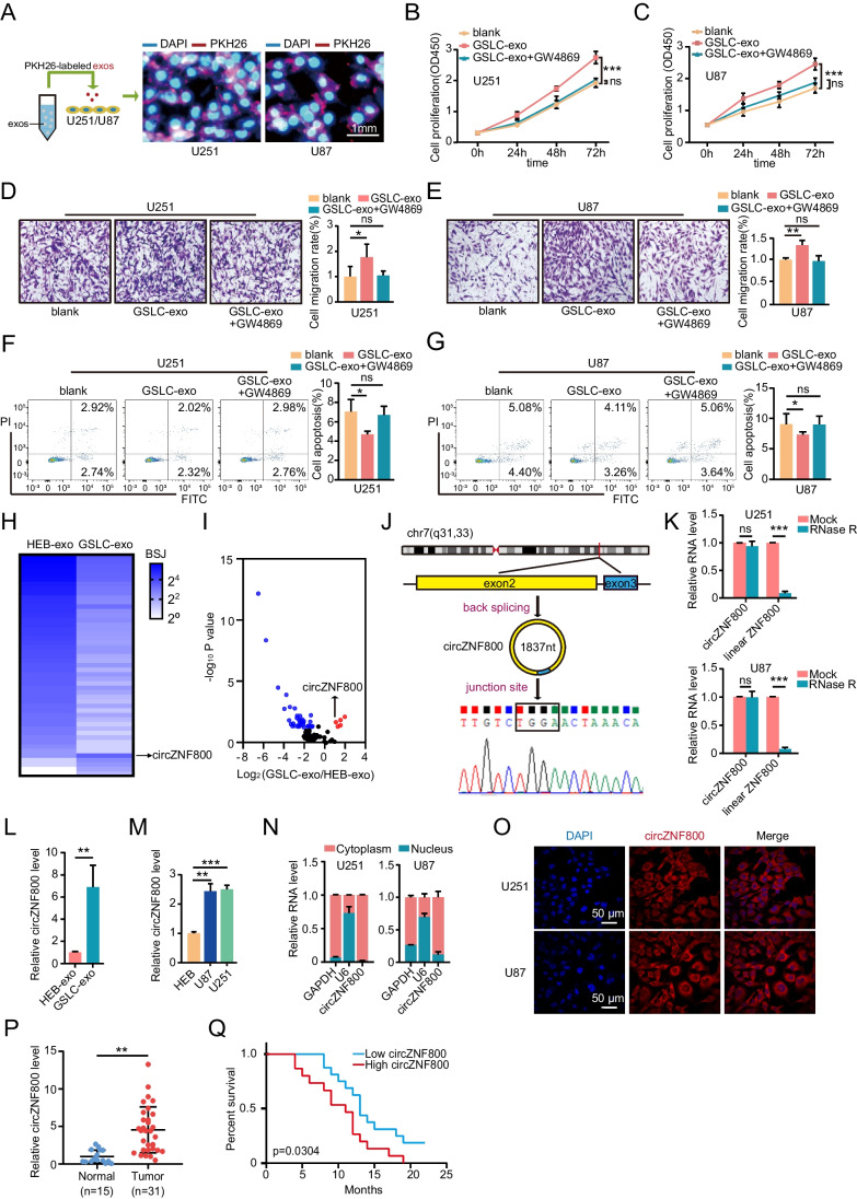
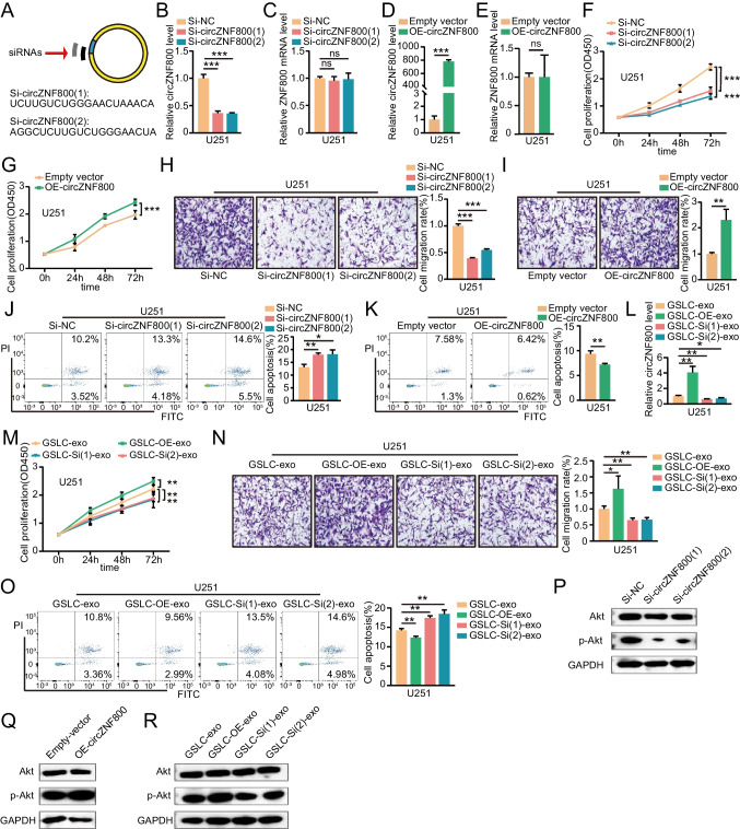
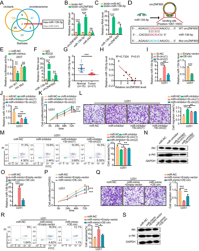
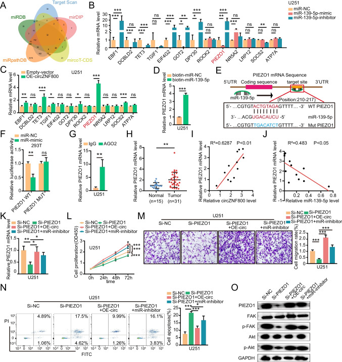
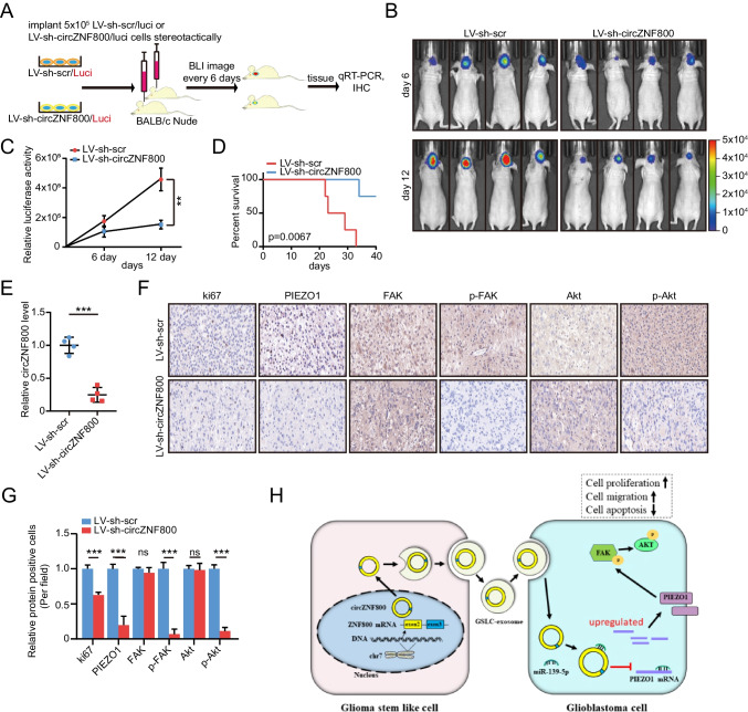
Exosomes play a crucial role in regulating crosstalk between tumor and tumor stem-like cells through their cargo molecules. Circular RNAs (circRNAs) have recently been demonstrated to be critical factors in tumorigenesis. This study focuses on the molecular mechanism by which circRNAs from glioma stem-like cell (GSLC) exosomes regulate glioblastoma (GBM) tumorigenicity. In this study, we validated that GSLC exosomes accelerated the malignant phenotype of GBM. Subsequently, we found that circZNF800 was highly expressed in GSLC exosomes and was negatively associated with GBM patients. CircZNF800 promoted GBM cell proliferation and migration and inhibited GBM cell apoptosis in vitro. Silencing circZNF800 could improve the GBM xenograft model survival rate. Mechanistic studies revealed that circZNF800 activated the PIEZO1/Akt signaling pathway by sponging miR-139-5p. CircZNF800 derived from GSLC exosomes promoted GBM cell tumorigenicity and predicted poor prognosis in GBM patients. CircZNF800 has the potential to serve as a promising target for further therapeutic exploration.

Keywords: Exosome; Glioma stem-like cell; PIEZO1; circZNF800; miR-139-5p.


Conflict of interest statement

The authors declare there are no potential competing interests.

Figures

Fig. 1
Fig. 1
CircZNF800 was overexpressed in GSLC-derived exosomes and correlated with poor patient prognosis. (A) Exosomes from GSLCs were labeled with PKH26 and then added to U251 and U87 cell cultures (PKH26-red, DAPI-blue). Scale bar, 1 mm. (B-C) CCK-8 assay was used to evaluate the viability of U251 and U87 cells treated with GSLC-exos. GW4869 is applied to inhibit exosomes. (D-E) Transwell experiments measured the migration of U251 and U87 cells treated with GSLC-exos. GW4869 is applied to inhibit exosomes. (F-G) Flow cytometry assays measured the apoptosis ratio of U251 and U87 cells co-cultured with GSLC-exos. GW4869 is applied to inhibit exosomes. (H) Heatmap showing the differential expression of circZNF800 in the HEB-exos and GSLC-exos. (I) Volcano plots illustrating differential changes of circRNAs in HEB-exos versus GSLC-exos samples. Blue and red dots represent significantly down-regulated and up-regulated circRNAs, respectively (J) Schematic illustration indicating the generation of circZNF800 from its host gene and junction site validation by Sanger sequencing. (K) QRT-PCR analysis of circZNF800 and ZNF800 mRNA after treatment with or without RNase R in U251 and U87 cells. (L) QRT-PCR analysis of the expression of circZNF800 in HEB-exos and GSLC-exos. (M) The expression level of circZNF800 in HEB cells and glioblastoma cells (U87 and U251) was measured by qRT-PCR. (N) Cytoplasm and nuclear fractions were used to detect the location of circZNF800. GAPDH was used as a cytoplasmic negative control. U6 was used as a nuclear negative control. (O) The localization of circZNF800 was detected by RNA FISH in U87 and U251 cells. Nuclei were stained with 4,6-diamidino-2-phenylindole (DAPI). CircZNF800 was labeled with Cyanine 3 (Cyy3) dye. Scale bar, 50 μm (P) QRT-PCR assay detected the expression of circZNF800 in glioblastoma tissues (n = 31) compared to normal tissues (n = 15). GAPDH was used as a control. (Q) The 31 glioblastoma samples were divided into high and low groups based on circZNF800 expression. Kaplan–Meier survival curve analysis showed the relationship between the expression circZNF800 and glioblastoma patient survival. Data are presented as the mean ± S.D. The P value was determined by Student's t test or Kaplan–Meier survival curve analysis. Significant results are presented as NS nonsignificant, *P < 0.05, **P < 0.01, or ***P < 0.001
Fig. 2
Fig. 2
CircZNF800 promotes glioblastoma proliferation and migration and inhibits glioblastoma apoptosis in vitro. (A) Schematic diagram of the siRNA sequences specifically targeting the circZNF800 junction. (B) The expression of circZNF800 analyzed by qRT-PCR in Si-circZNF800 U251 cells. GAPDH was used as a control. (C) The expression of ZNF800 mRNA in U251 cells treated with two independent siRNAs. GAPDH was used as a control. (D-E) QRT-PCR verified the expression of circZNF800 and ZNF800 mRNA after transduction of OE-circZNF800 plasmids in U251 cells. GAPDH was used as a control. (F-G) CCK-8 assay analysis the effect of circZNF800 knockdown and overexpression on U251 proliferation. (H-I) Transwell assay tested the effect of circZNF800 knockdown and overexpression on U251 cell migration. (J-K) Annexin‐V FITC/PI staining detected the effect of circZNF800 knockdown and overexpression on U251 apoptosis. (L) QRT-PCR analysis of circZNF800 expression in U251 cells after treatment with exosomes derived from circZNF800-overexpressing GSLC (GSLC-OE-exo) or knockdown GSLC (GSLC-Si(1)-exo and GSLC-Si(2)-exo) cells. (M) The proliferation of U251 cells treated with various exosomes (GSLC-OE-exo, GSLC-Si(1)-exo and GSLC-Si(2)-exo) or control were detected by CCK-8 assay. (N) Transwell assays were used to detect the migration ability of U251 cells treated with various exosomes (GSLC-OE-exo, GSLC-Si(1)-exo and GSLC-Si(2)-exo) or control. (O) Flow cytometry detected the apoptosis of U251 cells treated with GSLC-exos, GSLC-OE-exos, GSLC-Si(1)-exos or GSLC-Si(2)-exos. (P-R) Western blotting analysis revealed that p-Akt activation was regulated by circZNF800. U251 cells transfecting with (P) si-circZNF800(1) or si-circZNF800(2), (Q) overexpression circZNF800, (R) GSLC-OE-exos, GSLC-Si(1)-exos or GSLC-Si(2)-exos. Data are presented as the mean ± S.D. The P value was determined by Student's t test. Significant results are presented as NS nonsignificant, *P < 0.05, **P < 0.01, or ***P < 0.001
Fig. 3
Fig. 3
CircZNF800 functions as a sponge of miR-139-5p. (A) Venn diagram showing targets of circZNF800 predicted from circbank, circinteractome and StarBase. (B) Relative levels of circZNF800, miR-139-5p and miR-543 in U251 lysates were captured by the biotinylated probe circZNF800. GAPDH and U6 were used as controls. (C) Relative levels of circZNF800 and miR-139-5p in U251 lysates captured by the biotinylated probe of miR-139-5p. (D) Schematic of circZNF800 wild-type (WT) and mutant (Mut) luciferase reporter vectors. (E) Luciferase reporter gene assay to detect the interaction between circZNF800 and miR-139-5p. (F) RIP experiments were carried out in U251 cell extracts using an anti-AGO2 antibody. (G) The expression of miR-139-5p in GBM tissues and normal tissues by using qRT-PCR. U6 was used as a control. (H) Pearson correlation analysis of circZNF800 and miR-139-5p expression in GBM tissues (n = 12). (I) Expression level of miR-139-5p in U251 cells after transfection with si-circZNF800(1), si-circZNF800(2), or OE-circZNF800. (J) QRT-PCR was used to verify the efficiency of the miR-139-5p inhibitor was rescued by Si-circZNF800(1) and Si-circZNF800(2). (K-M) Detecting cell proliferation, migration and ratio of apoptosis of U251 treated miR-139-5p inhibitor alone or Si-circZNF800(1) and Si-circZNF800(2) respectively. (K) CCK-8 assays measured the migration ability of transfected U251 cells. (L) Transwell assays measured the migration ability of transfected U251 cells. (M) The apoptosis of treated U251 cells was detected by flow cytometry. (N) Western blotting was used to verify the activation level of p-Akt. (O) miR-139-5p expression in U251 cells transfected with miR-139-5p mimic alone or co-transfected with the OE-circZNF800 plasmid. (P) CCK-8 assay measured U251 cells transfected with miR-139-5p mimic alone or co-transfected with OE-circZNF800. (Q) The migration analysis of U251 cells transfected with miR-139-5p mimic alone or co-transfected with the OE-circZNF800 plasmid. (R) Annexin‐V FITC/PI staining was used to assess the apoptotic rates of U251 cells transfected with miR-139-5p mimic alone or co-transfected with the OE-circZNF800 plasmid. (S) The activation level of p-Akt was verified by western blotting. U251 cells transfected with miR-139-5p mimic or co-transfecting with the OE-circZNF800 plasmid. GAPDH was used as a control. Data are presented as the mean ± S.D. The P value was determined by Student's t test. Significant results are presented as NS nonsignificant, *P < 0.05, **P < 0.01, or ***P < 0.001
Fig. 4
Fig. 4
PIEZO1 is a direct target of miR-139-5p and is regulated by circZNF800. (A) The target genes of miR-139-5p predicted by TargetScan, miRDB, miRpathBD, microT-CDS, and miRDIP. (B-C) The mRNA expression levels of downstream genes targeted by miR-139-5p were measured by qRT-PCR in U251 cells treated with (B) miR-139-5p mimic or miR-139-5p inhibitor, (C) OE-circZNF800. (D) The RNA pulldown assay was performed with relative levels of PIEZO1 in U251 lysates captured by the biotinylated probe of miR-139-5p. (E) The schematic diagram shows the binding site of miR-139-5p and the PIEZO1 3'UTR. (F) Luciferase reporter gene assay to detect the interaction between circZNF800 and miR-139-5p. (G) The RIP assay detected the expression of PIEZO1 mRNA in U251 cell lysates using an anti-AGO2 antibody. IgG antibody was used as a control. (H) The expression of PIEZO1 mRNA in glioblastoma (n = 31) tissues and normal tissues (n = 15) was measured by qRT-PCR. (I) Pearson correlation analysis of PIEZO1 mRNA and circZNF800 expression in glioblastoma tissues (n = 12). (J) Pearson correlation analysis of PIEZO1 mRNA and miR-139-5p expression in glioblastoma tissues (n = 12). (K) The qRT-PCR assay detected the expression of PIEZO1 mRNA in U251 cells after transfection with Si-PIEZO1 alone or co-transfection with OE-circZNF800 or miR-139-5p inhibitor. (L) CCK-8 assay was performed to assess U251 cell growth ability after transfection with Si-PIEZO1 alone or co-transfection with OE-circZNF800 or miR-139-5p inhibitor. (M) Transwell assays were performed to assess the migration ability of U251 cells after transfection with Si-PIEZO1 alone or co-transfection with OE-circZNF800 or miR-139-5p inhibitor. (N) Flow cytometry analysis showed the apoptosis of U251 cells after transfection with Si-PIEZO1 alone or co-transfected with OE-circZNF800 or miR-139-5p inhibitor. (O) Western blotting analysis of the protein levels of PIEZO1, FAK, p-FAK, Akt and p-Akt in U251 cells. Data are presented as the mean ± S.D. The P value was determined by Student's t test. Significant results are presented as NS nonsignificant, *P < 0.05, **P < 0.01, or ***P < 0.001
Fig. 5
Fig. 5
Silencing circZNF800 can inhibit glioblastoma growth and metastasis in vivo. (A) The schematic diagram shows LV-sh-scr/luci and LV-sh-circZNF800/luci U251 cells (5 × 105 cells) orthotopic xenotransplantation in nude mice. (B) Bioluminescent images of nude mice. (C) Quantitative study of bioluminescence imaging signal intensity in nude mice. (D) Kaplan–Meier curve analysis showed the survival of xenograft models between LV-sh-scr and LV-sh-circZNF800 group. (E) The expression of circZNF800 was measured by qRT-PCR assay in xenograft tumors. (F) The expression of Ki-67, PIEZO1, FAK, p-FAK, Akt and p-Akt was examined by immunohistochemistry in xenograft tumors (400 ×). (G) Relative proteins (Ki-67, PIEZO1, FAK, p-FAK, Akt and p-Akt) positive cells in LV-sh-scr or LV-sh-circZNF800 cell-derived tissues were analyzed by IHC. (H) The schematic diagram shows how circZNF800 derived from GSLCs could promote tumorigenesis of GBM through circZNF800/PIEZO1/Akt axis. Data are presented as the mean ± S.D. The P value was determined by Student's t test or Kaplan–Meier survival curve analysis. Significant results are presented as NS nonsignificant, **P < 0.01, or ***P < 0.001

Similar articles

Cited by

References

    1. Meyer MA (2008) Malignant gliomas in adults. N Engl J Med 359(17):1850 author reply 1850 - PubMed
    1. Alexander BM, Cloughesy TF (2017) Adult Glioblastoma. J Clin Oncol 35(21):2402–2409 10.1200/JCO.2017.73.0119 - DOI - PubMed
    1. Lah TT, Novak M, Breznik B (2020) Brain malignancies: Glioblastoma and brain metastases. Semin Cancer Biol 60:262–273 10.1016/j.semcancer.2019.10.010 - DOI - PubMed
    1. Kaba SE, Kyritsis AP (1997) Recognition and management of gliomas. Drugs 53(2):235–244 10.2165/00003495-199753020-00004 - DOI - PubMed
    1. Sharanek A, Burban A, Laaper M, Heckel E, Joyal JS, Soleimani VD, Jahani-Asl A (2020) OSMR controls glioma stem cell respiration and confers resistance of glioblastoma to ionizing radiation. Nat Commun 11(1):4116 10.1038/s41467-020-17885-z - DOI - PMC - PubMed

MeSH terms

LinkOut - more resources