. 2024 Feb 9;10(6):eadi9284.
doi: 10.1126/sciadv.adi9284. Epub 2024 Feb 7.

NU6300 covalently reacts with cysteine-191 of gasdermin D to block its cleavage and palmitoylation

Affiliations

NU6300 covalently reacts with cysteine-191 of gasdermin D to block its cleavage and palmitoylation

Xueqin Jiang et al. Sci Adv. .

Abstract

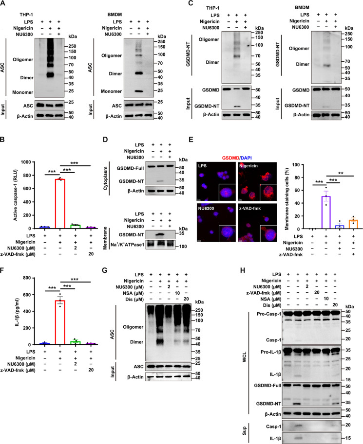
Gasdermin D (GSDMD) serves as a vital mediator of inflammasome-driven pyroptosis. In our study, we have identified NU6300 as a specific GSDMD inhibitor that covalently interacts with cysteine-191 of GSDMD, effectively blocking its cleavage while not affecting earlier steps such as ASC oligomerization and caspase-1 processing in AIM2- and NLRC4-mediated inflammation. On the contrary, NU6300 robustly inhibits these earlier steps in NLRP3 inflammasome, confirming a unique feedback inhibition effect in the NLRP3-GSDMD pathway upon GSDMD targeting. Our study reveals a previously undefined mechanism of GSDMD inhibitors: NU6300 impairs the palmitoylation of both full-length and N-terminal GSDMD, impeding the membrane localization and oligomerization of N-terminal GSDMD. In vivo studies further demonstrate the efficacy of NU6300 in ameliorating dextran sodium sulfate-induced colitis and improving survival in lipopolysaccharide-induced sepsis. Overall, these findings highlight the potential of NU6300 as a promising lead compound for the treatment of inflammatory diseases.


Figures

Fig. 1.
Fig. 1.. NU6300 inhibits pyroptosis.
(A) Experimental schematic of screening the active small molecules using LDH analysis. (B) THP-1 cells were stimulated with LPS (1 μg/ml) for 3 hours and then treated with different compounds (2 μM) for 40 min before they were induced with nigericin (10 μM) for 35 min, and the inhibition rate of cell death was determined by LDH analysis with a cutoff value of 75% inhibition. (C) Chemical structure of NU6300. (D and E) THP-1 cells and BMDMs were primed with LPS and incubated with NU6300 before stimulation with poly(deoxyadenylic-deoxythymidylic) acid [poly(dA:dT)] (500 ng/ml) (D) or flagellin (250 ng/ml) (E) for 6 hours, followed by LDH analysis of cell death. (F) THP-1 cells and BMDMs were treated with Pam3CSK4 (400 ng/ml) for 3 hours and incubated with NU6300 before stimulation with cytosolic LPS (1.5 μg/ml) overnight, followed by LDH analysis of cell death. (G) THP-1 cells and BMDMs were primed with LPS and incubated with NU6300 (2 μM), NSA (10 μM), or Dis (20 μM) before nigericin induction, followed by LDH analysis of cell death. (H) HT-29 cells were pretreated with or without NU6300 (2 μM) or NSA (10 μM) for 1 hour before stimulation with TNFα (25 ng/ml) (T), 400 nM SMAC mimetic (S), and 20 μM z-VAD-fmk (Z) for 24 hours, and cell viability was analyzed by CCK8 assay. (I) Kinetic analysis of PI uptake and cellular membrane permeability after treatment with NU6300 in THP-1 and BMDMs. (J) Transmission electron microscope observation of THP-1 cells morphology. Red arrows indicate representative organelles. Scale bars, 1 μm. Graphs showed means ± SEM, n = 3. Statistics were analyzed by one-way analysis of variance (ANOVA). *P < 0.05, **P < 0.01, and ***P < 0.001. ns, not significant.
Fig. 2.
Fig. 2.. NU6300 directly interacts with GSDMD.
(A) DARTS assay in LPS-primed THP-1 cells treated with dimethyl sulfoxide (DMSO) or NU6300 (1000, 100, and 10 μM). The lysate samples were digested with pronase (1:500, pronase-to-protein mass ratio) and analyzed by SDS–polyacrylamide gel electrophoresis and Coomassie brilliant blue staining. (B to E) Immunoblot assay to detect the CAP1 (B), NAPRT (C), TCP1 (D), and UMPS (E) proteins by DARTS analysis in LPS-primed THP-1 cells. (F to H) Immunoblot assay to GSDMD protein in THP-1 cells (F), BMDMs (G), and purified protein (H), and the relative density of GSDMD protein was obtained by normalization to the control group. (I and J) CETSA in LPS-primed THP-1 cell lysates (I) or TSA (J) in purified GSDMD protein. The samples were incubated with NU6300 (10 and 20 μM) at various temperatures, the relative density of GSDMD protein was normalized by the sample without heating, and ΔT1 and ΔT2 indicated the thermal shift of NU6300 (10 and 20 μM) as compared to DMSO control. (K) The binding of GSDMD with NU6300 was evaluated by BLI analysis, and the equilibrium binding signal (Req) was plotted against concentration of analyte. Data were presented as means ± SEM, n = 3. One-way ANOVA or two-way ANOVA was used. *P < 0.05, **P < 0.01, and ***P < 0.001. ns, not significant.
Fig. 3.
Fig. 3.. NU6300 directly binds to C191 of GSDMD.
(A) MS/MS spectra of the corresponding human GSDMD peptide FSLPGATCLQGEGQGHLSQK modified on C191 after GSDMD incubation with NU6300. (B) NU6300 (0.5, 1, and 2 μM) and NAC (500 μM) were preincubated for 1 hour before stimulation with nigericin (10 μM) from LPS-primed THP-1 cells for LDH assay. (C) HEK-293T cells were transfected with either GSDMD, p30, p30-C38A, p30-C56A, p30-C191A, or p30-C268A and treated with DMSO or NU6300 (2.5, 5, and 10 μM) before LDH analysis. (D) Inhibitory effect of NU6300 on pyroptosis was evaluated by transfection with GSDMD, p30, p30-C191A, p30-C38A/C56A/C268A, or p30-C38A/C56A/C191A/C268A in HEK-293T cells. (E) MST analysis by incubation of GSDMD protein (0.2 μM) with NU6300 or NU2. (F) Cell cytotoxicity of THP-1 cells was analyzed by stimulation with nigericin and then incubated with NU6300 or NU2 (0.5, 1, 2, and 4 μM). Data were expressed as means ± SEM, n = 3. Comparisons were calculated by one-way ANOVA. ***P < 0.001. ns, not significant.
Fig. 4.
Fig. 4.. NU6300 blocks the cleavage of GSDMD.
(A and B) ASC oligomerization detection in THP-1 cells or BMDMs by disuccinimidyl suberate cross-linking assays. Cells were primed with LPS (1 μg/ml), incubated with NU6300 (2 μM), and then transfected with poly(dA:dT) (500 ng/ml) (A) and flagellin (250 ng/ml) (B) for 6 hours. (C and D) The caspase-1 activity was measured by Caspase-Glo 1 reagent after AIM2 inflammasome (C) or NLRC4 inflammasome (D) activation in THP-1 cells and BMDMs. (E and F) GSDMD oligomerization and cleavage of GSDMD in THP-1 cells and BMDMs after AIM2 inflammasome (E) or NLRC4 inflammasome (F) activation. Data were expressed as means ± SEM, n = 3. Comparisons were calculated by one-way ANOVA. *P < 0.05, **P < 0.01, and ***P < 0.001. ns, not significant.
Fig. 5.
Fig. 5.. NU6300 blocks the palmitoylation and membrane translocation of GSDMD-N.
(A and B) THP-1 cells were primed with LPS (1 μg/ml) for 3 hours, followed by NU6300 (2 μM), and then transfected with poly(dA:dT) (500 ng/ml) (A) and flagellin (250 ng/ml) (B) for 6 hours, and cell lysates were treated with or without HA and subjected to the acyl-biotin exchange (ABE)–palmitoylation assay. (C) ABE-palmitoylation assay in HEK-293T cells transfected with p30 and incubated with NU6300 (5 μM). (D and E) Expression of GSDMD-NT in cell membrane and cytoplasm after treatment with NU6300 and transfected with poly(dA:dT) (D) or flagellin (E). (F and G) Expression of GSDMD-NT (p30) protein in cell membrane and cytoplasm (F) and the GSDMD-NT oligomerization (G) in HEK-293T cells transfected with p30 and incubated with NU6300 (5 μM). Data were expressed as means ± SEM, n = 3. Comparisons were calculated by one-way ANOVA. *P < 0.05, **P < 0.01, and ***P < 0.001. ns, not significant.
Fig. 6.
Fig. 6.. GSDMD inhibitors show a feedback inhibition on NLRP3 inflammasome.
(A) Oligomerization detection by disuccinimidyl suberate cross-linking assays. THP-1 cells and BMDMs were primed with LPS (1 μg/ml), followed by NU6300 (2 μM) treatment, and stimulated with nigericin (10 μM), and then the ASC oligomerization was detected. (B) LPS-primed THP-1 cells were incubated with NU6300 (2 μM) or z-VAD-fmk (20 μM) before stimulation with nigericin, and then the caspase-1 activity was measured by Caspase-Glo 1 reagent. (C) The cleavage and oligomerization of GSDMD were analyzed in LPS plus nigericin-activated THP-1 cells and BMDMs. (D) Membrane translocation assay in LPS and nigericin-activated THP-1 cells. (E) Confocal fluorescence microscope study for GSDMD membrane staining after incubation with NU6300 or z-VAD-fmk in LPS plus nigericin-activated THP-1 cells. Scale bars, 10 μm. (F) The release of IL-1β was detected by ELISA analysis. (G and H) LPS-primed THP-1 cells were incubated with NU6300 (2 μM), z-VAD-fmk (20 μM), NSA (10 μM), or Dis (20 μM) before stimulation with nigericin. The treated cells were analyzed for ASC oligomerization (G) and pro–caspase-1, GSDMD, and pro–IL-1β cleavage, the maturation and release of caspase-1 and IL-1β (H) by immunoblot of culture supernatants (Sup) or whole-cell lysate (WCL). Graphs showed means ± SEM, n = 3. Statistics were analyzed by one-way ANOVA. **P < 0.001 and ***P < 0.001.
Fig. 7.
Fig. 7.. NU6300 alleviates DSS-induced colitis and LPS-induced sepsis in mice.
(A) Flowchart of colitis mouse model induced by DSS (n = 6). (B) Daily loss weight of mice (n = 6). (C) The DAI was recorded (n = 6). (D) Colon length of mice (n = 6). (E) H&E staining to observe the pathological changes of colons. Scale bar, 200 μm. (F) The IL-1β and TNFα levels in colon tissues were determined by ELISA analysis (n = 6). (G) The protein expression of cleaved GSDMD in colons was detected by immunoblot assay (n = 3). (H) Flowchart of sepsis mouse model induced by LPS. (I) The survival curves were analyzed by using the log-rank (Mantel-Cox) test (n = 10). (J to L) Levels of IL-1β (J) and TNFα (K) in spleen were measured by ELISA, and spleen index (L) was analyzed after administration with LPS for 4 hours (n = 6). (M) Survival curves in wild-type and GSDMD−/− C57BL/6J mice after administration with or without NU6300 (10 mg/kg) for 1 hour and then after challenge with LPS (50 mg/kg) (n = 10). (N and O) Mice were pretreated with or without NU6300 (10 mg/kg) and then injected with LPS (50 mg/kg) for 4 hours, and the levels of IL-1β (N) and TNFα (O) in spleen were measured by ELISA (n = 6). (P) Cell cytotoxicity of BMDMs in wild-type and GSDMD−/− mice was analyzed by incubation with NU6300 and stimulation with nigericin (n = 3). Data were presented as means ± SEM, and one-way ANOVA was performed. *P < 0.05, **P < 0.01, and ***P < 0.001. ns, not significant.
Fig. 8.
Fig. 8.. The mechanism of NU6300 modulating pyroptosis.

References

    1. Shi J. J., Zhao Y., Wang K., Shi X. Y., Wang Y., Huang H. W., Zhuang Y. H., Cai T., Wang F. C., Shao F., Cleavage of GSDMD by inflammatory caspases determines pyroptotic cell death. Nature 526, 660–665 (2015). - PubMed
    1. Feng S., Fox D., Man S. M., Mechanisms of gasdermin family members in inflammasome signaling and cell death. J. Mol. Biol. 430, 3068–3080 (2018). - PubMed
    1. Ding J. J., Wang K., Liu W., She Y., Sun Q., Shi J. J., Sun H. Z., Wang D. C., Shao F., Pore-forming activity and structural autoinhibition of the gasdermin family. Nature 535, 111–116 (2016). - PubMed
    1. Kuang S. Y., Zheng J., Yang H., Li S. H., Duan S. Y., Shen Y. F., Ji C. N., Gan J. H., Xu X. W., Li J. X., Structure insight of GSDMD reveals the basis of GSDMD autoinhibition in cell pyroptosis. Proc. Natl. Acad. Sci. U.S.A. 114, 10642–10647 (2017). - PMC - PubMed
    1. A. Balasubramanian, L. Ghimire, A. Y. Hsu, H. Kambara, X. Liu, T. Hasegawa, R. Xu, M. Tahir, H. Yu, J. Lieberman, H. R. Luo, Palmitoylation of gasdermin D directs its membrane translocation and pore formation in pyroptosis. bioRxiv 2023.02.21.529402 [Preprint] (2023). 10.1101/2023.02.21.529402. - DOI

Publication types

MeSH terms

LinkOut - more resources