. 2024 Feb 9;10(6):eadk6722.
doi: 10.1126/sciadv.adk6722. Epub 2024 Feb 7.

Aligned cryogel fibers incorporated 3D printed scaffold effectively facilitates bone regeneration by enhancing cell recruitment and function

Affiliations

Aligned cryogel fibers incorporated 3D printed scaffold effectively facilitates bone regeneration by enhancing cell recruitment and function

Yuxuan Wei et al. Sci Adv. .

Abstract

Reconstructing extensive cranial defects represents a persistent clinical challenge. Here, we reported a hybrid three-dimensional (3D) printed scaffold with modification of QK peptide and KP peptide for effectively promoting endogenous cranial bone regeneration. The hybrid 3D printed scaffold consists of vertically aligned cryogel fibers that guide and promote cell penetration into the defect area in the early stages of bone repair. Then, the conjugated QK peptide and KP peptide further regulate the function of the recruited cells to promote vascularization and osteogenic differentiation in the defect area. The regenerated bone volume and surface coverage of the dual peptide-modified hybrid scaffold were significantly higher than the positive control group. In addition, the dual peptide-modified hybrid scaffold demonstrated sustained enhancement of bone regeneration and avoidance of bone resorption compared to the collagen sponge group. We expect that the design of dual peptide-modified hybrid scaffold will provide a promising strategy for bone regeneration.


Figures

Fig. 1.
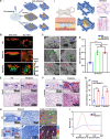
Fig. 1.. ACFs incorporated 3D PS effectively facilitates bone regeneration by enhancing cell recruitment and function.
(A) Fabrication procedure of ACFs incorporated into a 3D PS and cell recruitment methods of PS/ACFs in the cranial bone defect. (B) PS/ACFs functionalized with KP and QK peptides facilitate critical-sized cranial bone defect repair in rats. Red arrows indicate the cell recruitment, blue arrows show the orientation of the newly formed collagenous fibers, and orange arrows indicate the osteogenic differentiation. BMSC, bone marrow mesenchymal stromal cell.
Fig. 2.
Fig. 2.. Morphological features of 3D printed scaffold embedded without/with ACFs.
(A) Schematic illustrating the preparation procedures of ACFs incorporated 3D PS by 3D printing in combination with freeze-casting. (B) Gross view of PS and PS/ACFs before and after lyophilization. (C) SEM images showing the top view and cross section of the PS and PS/ACFs. (D) False color images of ACFs indicate the angle mapping of fiber orientations. (E) Angle distribution of formed cryogel fibers. (F) Distribution of cryogel fibers’ diameter. Oct, optimal cutting temperature compound; UV, ultraviolet.
Fig. 3.
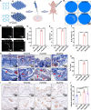
Fig. 3.. The in vitro cell seeding efficiency and in vivo cell recruitment ability of the ACFs incorporated 3D PS.
(A) (i) Schematic illustrating the in vitro cell seeding and adhesion on the PS, PS/RCFs, and PS/ACFs. (ii) Schematic illustrating the in vivo cell recruitment and tissue ingrowth induced by PS/ACFs by a subcutaneous implantation model in rats. (B and C) Confocal microscope 3D images and SEM images showing HUVECs adhesion and distribution on the PS, PS/RCFs, and PS/ACFs after 24 hours of cell seeding. (D) Quantification of attached HUVECs on the PS, PS/RCFs, and PS/ACFs after 24 hours of culture. (E to G) H&E staining and semi-quantitative analysis exhibit the cell infiltration and tissue ingrowth of PS and PS/ACFs after 1 and 2 weeks of subcutaneous implantation. (H) Trichrome staining discovers the newly formed collagenous fiber orientation of the ingrowth soft tissue in the PS and PS/ACFs groups after 2 weeks of operation. The false color images indicate the orientation of collagenous fiber. (I) Angle distribution of the newly formed collagenous fibers after 2 weeks of operation. ***P < 0.001 and ****P < 0.0001. ns, not significant.
Fig. 4.
Fig. 4.. Pro-osteogenic properties of the KP peptide-functionalized PS.
(A) Schematic illustrating the preparation procedures and verification of pro-osteogenic property of the KP peptide-functionalized PS. The ectopic osteogenesis model was used by subcutaneous implantation of rBMSCs-seeding scaffolds in nude mice (n = 5). (B) Micro-CT 3D reconstruction images exhibited ectopic new bone formation in the PS, PS-KP250 (250 μg/ml), PS-KP500 (500 μg/ml), and PS-KP750 (750 μg/ml) group after 4 weeks of operation. (C) Micro-CT axial, sagittal, and coronal views of scaffold area after 4 weeks of operation. (D to G) Regenerated bone volume (BV/TV, %), bone surface (BS/TS, %), Tb.N (1/100 mm), and Tb.Th (μm) after 4 weeks of operation. (H) Trichrome staining of the decalcified scaffold area and surrounding tissue of PS, PS-KP250, PS-KP500, and PS-KP750 groups after 4 weeks of operation. (I and J) OPN immunohistochemical staining of the decalcified scaffold area of PS, PS-KP250, PS-KP500, and PS-KP750 groups after 4 weeks of operation and semi-quantitative analysis. *P < 0.05, **P < 0.01, ***P < 0.001, and ****P < 0.0001. IOD, integral optical density.
Fig. 5.
Fig. 5.. Pro-angiogenic properties of QK peptide-functionalized PS/ACFs.
(A) Schematic illustrating the preparation procedures and verification of pro-angiogenic property of the QK peptide-functionalized PS/ACFs. The subcutaneous implantation of QK peptide-functionalized PS/ACFs in rats was used to evaluate its pro-angiogenic properties (n = 4). (B) Trichrome staining of the scaffold area and surrounding tissues of PS/ACFs, PS/ACFs-QK100 (100 μg/ml), PS/ACFs-QK300 (300 μg/ml), and PS/ACFs-QK500 (500 μg/ml) groups after 2 weeks of operation. (C) Quantification of the number of newly formed blood vessels in the PS/ACFs, PS/ACFs-QK100, PS/ACFs-QK300, and PS/ACFs-QK500 groups after 2 weeks of operation. (D) CD31 immunohistochemical staining of the scaffold area and surrounding tissues in the PS/ACFs, PS/ACFs-QK100, PS/ACFs-QK300, and PS/ACFs-QK500 groups after 2 weeks of operation. (E) Quantification of the area of newly formed blood vessels in the PS/ACFs, PS/ACFs-QK100, PS/ACFs-QK300, and PS/ACFs-QK500 group after 2 weeks of operation. *P < 0.05 and ***P < 0.001.
Fig. 6.
Fig. 6.. The bone pro-regenerative ability of KP peptide- and QK peptide-functionalized PS/ACFs in rat critical cranial bone defect model.
(A) Schematic illustrating the implantation of experimental scaffold (PS-KP500/ACFs-QK500) and collagen sponge (positive control) to a critical-sized cranial bone defect (φ 8 mm) in rats (n = 5). (B) X-ray raw images of the control, collagen sponge, and PS-KP500/ACFs-QK500 groups after 4 and 8 weeks of operation. (C and D) Micro-CT 3D reconstruction images and coronal views of the control, collagen sponge, and PS-KP500/ACFs-QK500 groups after 4 and 8 weeks of operation. (E and F) Regenerated bone volume (BV/TV, %) and surface coverage (%) of the control, collagen sponge, and PS-KP500/ACFs-QK500 groups after 4 and 8 weeks of operation. *P < 0.05, **P < 0.01, and ***P < 0.001.
Fig. 7.
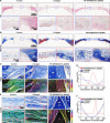
Fig. 7.. Histological observations of bone regeneration after 4 weeks of treatment.
(A and B) H&E and trichrome staining of the decalcified cranial bone of the control, collagen sponge, and PS-KP500/ACFs-QK500 groups after 4 weeks of operation (n = 5). (C and E) Trichrome staining of newly formed soft tissues in the edge and center area of the control, collagen sponge, and PS-KP500/ACFs-QK500 groups after 4 weeks of operation. The false color images indicate the orientation of the regenerated soft tissue. (D and F) Angle distribution of the regenerated collagenous fibers in the edge and center of the regenerated soft tissue after 4 weeks of operation. NB, new bone; C, collagen sponge; PS, printed scaffold.
Fig. 8.
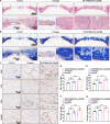
Fig. 8.. Histological observations of bone regeneration after 8 weeks of treatment.
(A and B) H&E and trichrome staining of the decalcified cranial bone of the control, collagen sponge, and PS-KP500/ACFs-QK500 groups after 8 weeks of operation (n = 5). (C) CD31, OCN, OPN, and RUNX2 immunohistochemical staining of the regenerated bone tissue of the control, collagen sponge, and PS-KP500/ACFs-QK500 groups after 8 weeks of operation. (D to G) Quantification of integral OD of expressed CD31, OCN, OPN, and RUNX2 in the control, collagen sponge, and PS-KP500/ACFs-QK500 groups after 8 weeks of operation. *P < 0.05, **P < 0.01, and ****P < 0.0001.

References

    1. Gillman C. E., Jayasuriya A. C., FDA-approved bone grafts and bone graft substitute devices in bone regeneration. Mater. Sci. Eng. C Mater. Biol. Appl. 130, 112466 (2021). - PMC - PubMed
    1. Elsalanty M. E., Genecov D. G., Bone grafts in craniofacial surgery. Craniomaxillofac. Trauma Reconstr. 2, 125–134 (2009). - PMC - PubMed
    1. Berretta S., Evans K., Ghita O., Additive manufacture of PEEK cranial implants: Manufacturing considerations versus accuracy and mechanical performance. Mater. Des. 139, 141–152 (2018).
    1. Takizawa T., Nakayama N., Haniu H., Aoki K., Okamoto M., Nomura H., Tanaka M., Sobajima A., Yoshida K., Kamanaka T., Ajima K., Oishi A., Kuroda C., Ishida H., Okano S., Kobayashi S., Kato H., Saito N., Titanium fiber plates for bone tissue repair. Adv. Mater. 30, 1703608 (2018). - PubMed
    1. I. Yadroitsava, A. Du Plessis, I. Yadroitsev, Bone regeneration on implants of titanium alloys produced by laser powder bed fusion: A review, in Titanium for Consumer Applications (Elsevier, 2019), chap. 12, pp. 197–233.

Publication types