. 2024 Feb 7;10(1):10.
doi: 10.1038/s41514-023-00130-4.

Mitochondrial function in peripheral blood cells across the human lifespan

Affiliations

Mitochondrial function in peripheral blood cells across the human lifespan

Johannes K Ehinger et al. NPJ Aging. .

Abstract

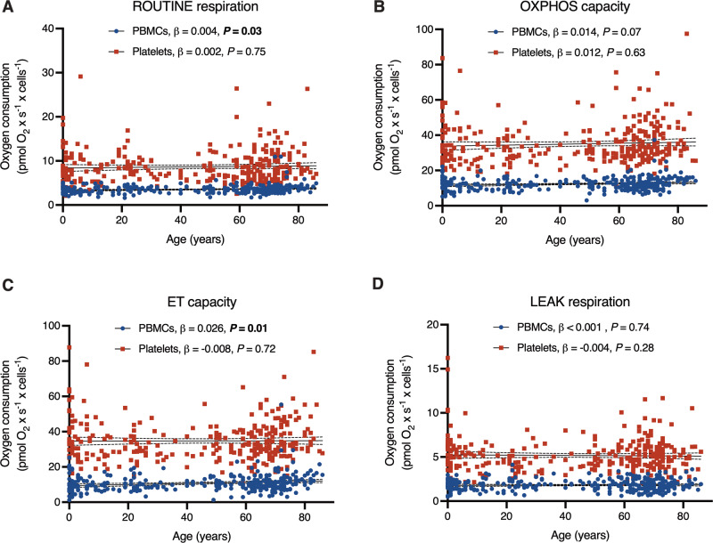
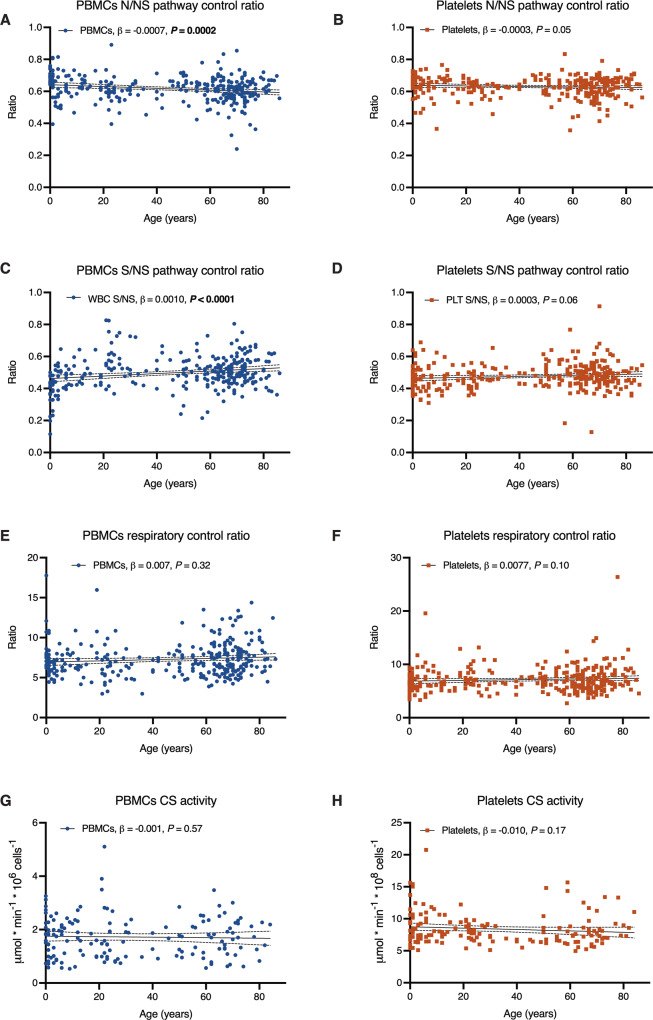
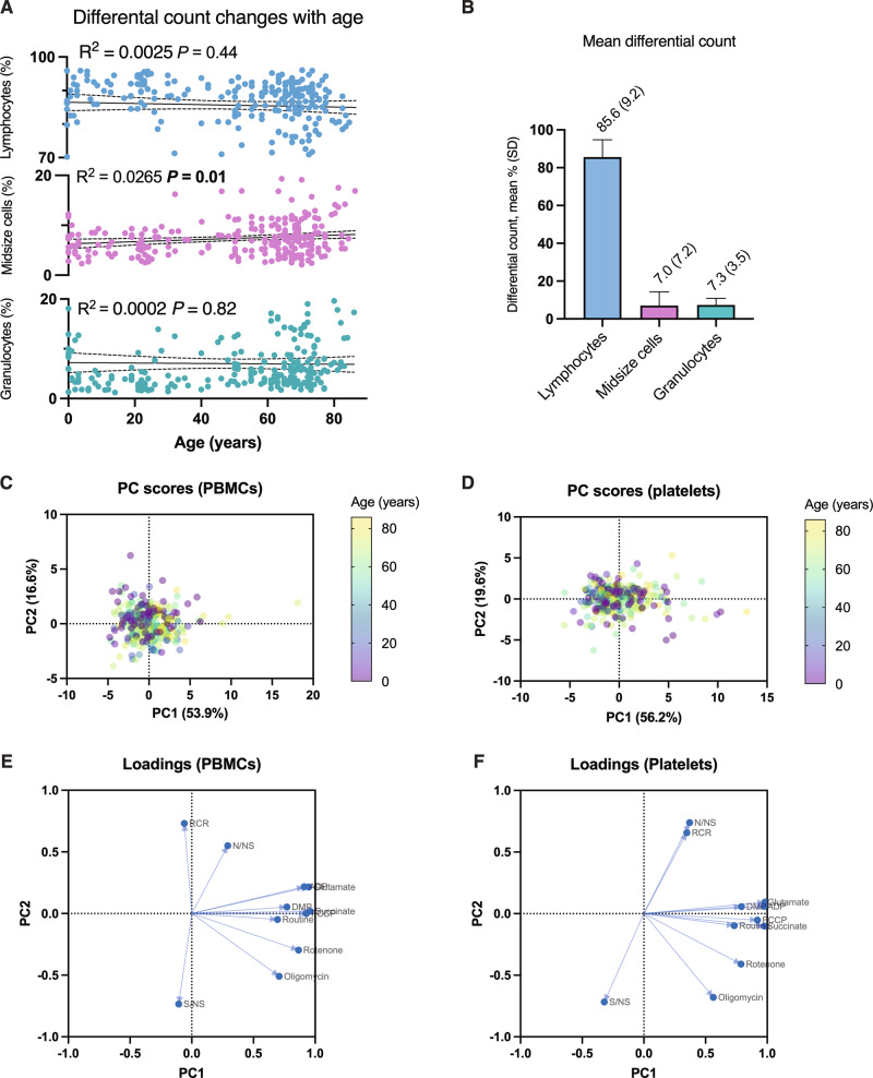
Mitochondrial dysfunction is considered a hallmark of aging. Up to now, a gradual decline of mitochondrial respiration with advancing age has mainly been demonstrated in human muscle tissue. A handful of studies have examined age-related mitochondrial dysfunction in human blood cells, and only with small sample sizes and mainly in platelets. In this study, we analyzed mitochondrial respiration in peripheral blood mononuclear cells (PBMCs) and platelets from 308 individuals across the human lifespan (0-86 years). In regression analyses, with adjustment for false discovery rate (FDR), we found age-related changes in respiratory measurements to be either small or absent. The main significant changes were an age-related relative decline in complex I-linked respiration and a corresponding rise of complex II-linked respiration in PBMCs. These results add to the understanding of mitochondrial dysfunction in aging and to its possible role in immune cell and platelet senescence.


Conflict of interest statement

J.E., E.Å.F., M.K., and E.E. have equity interests in and/or have received salary support from Abliva AB (formerly NeuroVive Pharmaceutical AB), a public company developing pharmaceuticals in the field of mitochondrial medicine. The remaining authors declare no competing interests.

Figures

Fig. 1
Fig. 1. Cell count-normalized respiration as a function of age, with adjustment for confounders.
(AD) Plots depict simple linear regression for each respiratory parameter (y-axes) depending on age (x-axes); the slope is depicted as a straight line with its 95% confidence interval as a dotted line (complete data in Supplementary Table 3). The age effect, and its P-value, adjusted for sex and health status in a multiple regression (MR) model, is displayed in the respective graph legend. None of the age effects were classified as true discoveries after adjustment for the false discovery rate. The overall MR model was significant for routine respiration (R2 = 0.027, P = 0.04), OXPHOS capacity (R2 = 0.036, P = 0.01), and ET capacity (R2 = 0.047, 0.002) for PBMCs; none of the MR models were significant for platelets (complete MR data in Supplementary Table 4).
Fig. 2
Fig. 2. Ratios and CS activity as a function of age, with adjustment for confounders.
Plots depict simple linear regression for each ratio (AF) and citrate synthase (CS, GH) activity (y-axes) depending on age (x-axes); the slope is depicted as a straight line with its 95% confidence interval as a dotted line (complete data in Supplementary Table 3). The age effect, and its P-value, adjusted for sex and health status in a multiple regression (MR) model, is displayed in the respective graph legend. Age effect P-values in bold were significant and classified as true discoveries after adjustment for false discovery rate. The overall MR model was significant for the N/NS pathway control ratio (R2 = 0.047, P = 0.002) and S/NS pathway control ratio (R2 = 0.086, P < 0.0001) for PBMCs; none of the MR models were significant for platelets (complete MR data in Supplementary Table 5).
Fig. 3
Fig. 3. Differential count and principal component analysis.
Linear regression of differential count (%) depending on age (A) for lymphocytes, midsize cells, and granulocytes (n = 262; slopes in Supplementary Table 6). 12, 1, and 8 data points, respectively, were removed from the lymphocyte, midsize cell, and granulocyte plots to compress the figure and enhance legibility. Mean differential count (%) and standard deviation (SD) for the same sample (B). Principal component (PC) scores for all respiratory measurements and derived ratios (Supplementary Tables 1 and 2) for PBMCS (C) and platelets (D) labeled by age. Corresponding loading plots for all parameters, for PBMCs (E) and platelets (F) (table of loadings in Supplementary Table 5).

References

    1. Harman D. Aging: a theory based on free radical and radiation chemistry. J. Gerontol. 1956;11:298–300. doi: 10.1093/geronj/11.3.298. - DOI - PubMed
    1. Zhang Y, et al. Mice deficient in both Mn superoxide dismutase and glutathione peroxidase-1 have increased oxidative damage and a greater incidence of pathology but no reduction in longevity. J. Gerontol. Ser. A. 2009;64:1212–1220. doi: 10.1093/gerona/glp132. - DOI - PMC - PubMed
    1. Van Raamsdonk JM, Hekimi S. Superoxide dismutase is dispensable for normal animal lifespan. Proc. Natl Acad. Sci. USA. 2012;109:5785–5790. doi: 10.1073/pnas.1116158109. - DOI - PMC - PubMed
    1. Sun N, Youle RJ, Finkel T. The mitochondrial basis of aging. Mol. Cell. 2016;61:654–666. doi: 10.1016/j.molcel.2016.01.028. - DOI - PMC - PubMed
    1. Bratic A, Larsson NG. The role of mitochondria in aging. J. Clin. Investig. 2013;123:951–957. doi: 10.1172/JCI64125. - DOI - PMC - PubMed