. 2024 Feb 7;15(1):1164.
doi: 10.1038/s41467-024-45396-8.

Structural basis for the oligomerization-facilitated NLRP3 activation

Affiliations

Structural basis for the oligomerization-facilitated NLRP3 activation

Xiaodi Yu et al. Nat Commun. .

Abstract

The NACHT-, leucine-rich-repeat-, and pyrin domain-containing protein 3 (NLRP3) is a critical intracellular inflammasome sensor and an important clinical target against inflammation-driven human diseases. Recent studies have elucidated its transition from a closed cage to an activated disk-like inflammasome, but the intermediate activation mechanism remains elusive. Here we report the cryo-electron microscopy structure of NLRP3, which forms an open octamer and undergoes a ~ 90° hinge rotation at the NACHT domain. Mutations on open octamer's interfaces reduce IL-1β signaling, highlighting its essential role in NLRP3 activation/inflammasome assembly. The centrosomal NIMA-related kinase 7 (NEK7) disrupts large NLRP3 oligomers and forms NEK7/NLRP3 monomers/dimers which is a critical step preceding the assembly of the disk-like inflammasome. These data demonstrate an oligomeric cooperative activation of NLRP3 and provide insight into its inflammasome assembly mechanism.


Conflict of interest statement

The authors declare the following competing interests: X.Y., R.Ma., R.Mi., D.C., B.V.S., K.G., J.S., B.P., Y.Y., G.J.T., L.P., A.B., D.O., N.V.O., and S.S. are employees of Johnson & Johnson Innovative Medicine. The remaining authors declare no competing interests.

Figures

Fig. 1
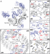
Fig. 1. Cryo-EM structure of open NLRP3ΔPYD/+ATP/-MCC950 octamer.
A Domain organization of human NLRP3 with PYD domain truncated. B Different views of cryo-EM map of NLRP3ΔPYD/+ATP/-MCC950. The NACHT and LRR domains were in slate and gray, respectively, with the key interfaces between protomers highlighted. One NLRPΔPYD protomer was highlighted. 2D classes from cryo-EM images of the sample were shown at the bottom. C Structural overlay of open NLRP3ΔPYD/+ATP/-MCC950 to the closed NLRP3 (Left, PDB ID: 7LFH) or activated NEK7/NLRP3 (Right, PDB ID: 8EJ4). The LRR region and NEK7 were colored in gray and orange, respectively. The NACHT domains were in cyan, slate, or green for the closed, open, or activated states, respectively. D Zoom-in views of NACHT conformational changes. NBD and HD1 were in orange and cyan or magenta and slate for the closed or open states, respectively. WHD and HD2 were in light and dark green, respectively. The nucleotide and MCC950 from the closed NLRP3 were shown as sticks. The protein structures were shown as cartoon. Subdomain reorganization upon activation was highlighted by arrows.
Fig. 2
Fig. 2. Interfaces in NLRP3ΔPYD/+ATP/-MCC950 octamer assembly.
A Interfaces in NLRP3ΔPYD/+ATP/-MCC950 octamer assembly. Squares indicated the locations of the interfaces. BE Zoomed-in views of Back-Back, Head-Face, Tail-Tail, or Face-Face interfaces, respectively. The main and side chains of residues at the interfaces were shown as cartoon and sticks, respectively, and following the color code in Fig. 1A.
Fig. 3
Fig. 3. Purposed NLRP3 activation mechanism.
A Schematic representation of the arrangement of the oligomerization transition from the closed to the open cages. NACHT was not shown. The Back-Back, Face-Face, Head-Face, and Tail-Tail interactions were highlighted using magenta, gray, pink, and yellow spheres, respectively. NLRP3 dimers with Back-Back interactions was highlighted as one unit. B Structural overlays of NLRP3 closed cage (PDB ID: 7LFH) and open octamer (this study) at face-face interface using one protomer as the reference. NACHT were colored in slate. LRR were in red or gray for closed cage or open octamer, respectively. C Structural overlays of NLRP3 closed (red, PDB ID: 7LFH) and open (slate and gray, this study) cages. LRRs were highlighted. D Schematic representation of the proposed activation mechanism at the NACHT domain in the octamer. Note, two consecutive units were shown.
Fig. 4
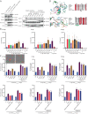
Fig. 4. Interfaces in the open octamer are critical in the NLRP3 activation and assembly.
A Mutations at the interfaces did not impair NLRP3 expression level. The component proteins were detected using Western blots. These experiments were repeated twice. B Cellular assay showing induction of IL-1β signaling upon transfection with 2.5, 5, or 10 ng of WT or mutant FL NLRP3 vectors. EV, empty vector control. Bars indicate the mean, and the data points from six independent experiments are shown. C Structural and sequence alignments around the catalytic surface around Asp363 (top) and the Loop504-517 region (lower), respectively. Closed (PDB ID: 7LFH), open (this study), activated (PDB ID: 8EJ4) NLRP3, NLRC4 (PDB ID: 8FW2), and NAIP (PDB ID: 8FVU) were shown as cartoon and in cyan, slate, light green, pink, and orange, respectively. The adjacent activated NLRP3 protomer was colored in dark green. Side chains of selected residues were shown in sticks in the structural alignments and highlighted with filled gray boxes in the sequence alignments. D Fluorescent mCherry measurements in HEK293TASC-mCherry cells transfected with empty vector (EV), NLRP3 WT, or mutants, with/without Nigericin treatment. Two cell images with scale bars were shown as reference. Bars indicate the mean, and the data points from three independent experiments are shown. Source data are provided as a Source Data file.
Fig. 5
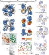
Fig. 5. Cryo-EM structures of NLRP3ΔPYD/+ATP/+MCC950 in complex with NEK7.
A, B Cryo-EM maps of closed NLRP3ΔPYD/+ATP/+MCC950 hexamer and NEK7/NLRP3ΔPYD/+ATP/+MCC950 dimer, respectively. Domain organization of NEK7 was shown. The NACHT, LRR, NEK7-N-lobe, and NEK7-C-lobe were colored in slate, gray, red, and orange, respectively. One protomer was highlighted, respectively. C Structure of closed NLRP3ΔPYD/+ATP/+MCC950 protomer within the hexamer (Left) and structural overlay of closed NLRP3ΔPYD/+ATP/+MCC950 hexamer and NEK7/NLRP3ΔPYD/+ATP/+MCC950 dimer at MCC950 binding site (Right). ATP and MCC950 were shown as sticks. NACHT subdomains NBD, HD1, WHD, and HD2 were colored in orange, slate, light green, and dark green, respectively. Loop617-628 was highlighted. D From top to down, structure of NEK7/NLRP3ΔPYD/+ATP/+MCC950 dimer, docking of NEK7 to the concave sides of LRRs in the closed hexamer (this study), FL cage (PDB ID: 7LFH) and open octamer (this study). The Back-Back, Face-Face, Head-Face, and Tail-Tail interactions were highlighted using magenta, gray, pink, and yellow spheres, respectively. The NACHT, LRR, and NEK7 were colored in slate, gray, and orange, respectively.
Fig. 6
Fig. 6. Proposed NLRP3 activation and uni- or bi- directional inflammasome assembly mechanisms.
The priming step increases NLRP3 levels, which leads to NLRP3 oligomerization. The activation of NLRP3 occurs through ATP hydrolysis and oligomer reorganization, forming an open octamer. These closed or open oligomerization states are favorable for trafficking on microtubules to the MTOC. NEK7 then dissociates the oligomer into NEK7/NLRP3 binary complexes in equilibrium with dimer and monomer. The open NEK7/NLRP3 allows for uni- or bi- directional inflammasome assembly as indicated by the gray arrows, while the closed NEK7/NLRP3 can also be passively activated by the open NEK7/NLRP3. MCC950 inhibits NLRP3’s ATPase hydrolysis activity, binds to all NACHT subdomains, and locks it in a closed conformation. The closed cage, NEK7/NLRP3 monomer, and the fully activated NLRP3 inflammasome were derived from PDB ID: 7LFH, 6NPY, and 8EJ4, respectively. The dashed arrows illustrate how MCC950 or NEK7’s early dissociation of the closed cage prevents NLRP3 activation.

References

    1. Eigenbrod T, Dalpke AH. Bacterial RNA: an underestimated stimulus for innate immune responses. J. Immunol. 2015;195:411–418. doi: 10.4049/jimmunol.1500530. - DOI - PubMed
    1. Inoue M, Shinohara ML. The role of interferon-beta in the treatment of multiple sclerosis and experimental autoimmune encephalomyelitis—in the perspective of inflammasomes. Immunology. 2013;139:11–18. doi: 10.1111/imm.12081. - DOI - PMC - PubMed
    1. Sharif H, et al. Structural mechanism for NEK7-licensed activation of NLRP3 inflammasome. Nature. 2019;570:338–343. doi: 10.1038/s41586-019-1295-z. - DOI - PMC - PubMed
    1. Ohto U, et al. Structural basis for the oligomerization-mediated regulation of NLRP3 inflammasome activation. Proc. Natl Acad. Sci. USA. 2022;119:e2121353119. doi: 10.1073/pnas.2121353119. - DOI - PMC - PubMed
    1. Hochheiser IV, et al. Structure of the NLRP3 decamer bound to the cytokine release inhibitor CRID3. Nature. 2022;604:184–189. doi: 10.1038/s41586-022-04467-w. - DOI - PubMed

MeSH terms