. 2024 Feb 7;14(1):3146.
doi: 10.1038/s41598-024-53661-5.

Anti-aquaporin-4 immune complex stimulates complement-dependent Th17 cytokine release in neuromyelitis optica spectrum disorders

Collaborators, Affiliations

Anti-aquaporin-4 immune complex stimulates complement-dependent Th17 cytokine release in neuromyelitis optica spectrum disorders

Shuhei Nishiyama et al. Sci Rep. .

Abstract

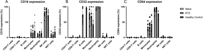
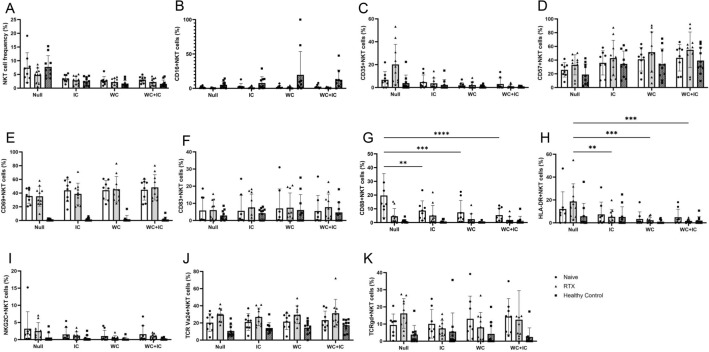
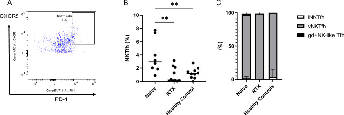
Proinflammatory cytokines, such as (IL: interleukin) IL-6 and IL-17A, and complement fixation are critical in the immunopathogenesis of neuromyelitis optica spectrum disorders (NMOSD). Blocking the IL-6 receptor or the C5 complement pathway reduces relapse risk. However, the role of interleukin (IL)-6 and complement in aquaporin-4 (AQP4) autoimmunity remains unclear. To investigate the role of the anti-AQP4 immunoglobulin (AQP4-IgG)/AQP4 immunocomplex on the induction and profile of ex vivo cytokine and surface marker expression in peripheral blood mononuclear cells (PBMC) culture. Isolated PBMCs obtained from 18 patients with AQP4-IgG-seropositive-NMOSD (8 treatment-naive, 10 rituximab-treated) or ten healthy controls were cultured with AQP4-immunocomplex with or without complement. Changes in PBMC surface markers and cytokine expression were profiled using flow cytometry and ELISA. PBMCs derived from treatment-naive NMOSD patients stimulated with a complex mixture of serum complement proteins produced significant elevations of IL-17A and IL-6. Rituximab-treated patients also exhibited higher IL-6 but not IL-17A release. IL-6 and IL-17A elevations are not observed without complement. Co-stimulation of PBMCs with AQP4-IgG/AQP4 immunocomplex and complement prompts a Th17-biased response consistent with the inflammatory paradigm observed in NMOSD. A possible inflammation model is proposed via antigen-specific autoreactive peripheral blood cells, including NK/NKT cells.


Conflict of interest statement

The authors declare no competing interests.

Figures

Figure 1
Figure 1
AQP4-immunocomplex kinetics and NKT cells in NMOSD. PBMCs from NMOSD patients and healthy controls (HC) were incubated with AQP4 protein + AQP4-IgG-containing serum (A), and analyzed by flow cytometry. After excluding doublet and dead cells, AQP4 positive cells were gated from lymphocytes (B). When cultured with the immunocomplexes for 8 h, the group using NMOSD patient-derived PBMCs and AQP4 immunocomplexes detects an AQP4-derived signal ((B) top row). In contrast, this signal cannot be detected in the group using control IgG + AQP4 protein ((B) middle row) or healthy control PBMCs ((B) bottom row). The difference between AQP4-positive lymphocytes with (red dots) or without (blue dots) permeabilization was calculated to detect internalized AQP4. AQP4-positive cells were increased in time-dependent manner (C). PBMCs treated with Control-IgG and AQP4 protein did not show AQP4-positive signals. There was little difference between those with and without permeabilization. Majority of the AQP4-positive cells were NKT cells (D), and among them, almost all the NKT cell were variant NKT cells (E,F). PD-1-positive CXCR5-positive subsets were extracted from CD3 + CD56 + CD11b-CD14-CD20-CD66b-NKT cells. + P with permeabilization, -P without permeabilization, AQP4-IC AQP4 protein/AQP4-IgG immunocomplexes, Ctrl-IgG Control-IgG, HC healthy controls, RTX rituximab-treated patients. NMOSD-Naive (n = 8), NMOSD-rituximab treated (n = 10), and healthy controls (n = 10). *p < 0.05; **p < 0.01; ***p < 0.001; ****p < 0.0001.
Figure 2
Figure 2
The expression levels of Fc gamma receptors in PBMCs. The expression levels of CD16 (A), CD32 (B), and CD64 (C) were obtained by flow cytometry. NK cells were predominantly CD16 expressing. NKT cells expressed CD16 as well but showed decreased expression in the NMOSD group. Neither CD4 + T cells nor CD8 + T cells expressed Fcɤ receptors. CD32 was positive on B cells, but CD16 and CD64 were not. We couldn’t detect any B cells from all rituximab-treated NMOSD patients. Most of the monocytes had CD32 signals in NMOSD groups. Some monocytes expressed CD16, thought to be either intermediate or non-classical monocytes. Neutrophils were expressing all Fcɤ receptors.
Figure 3
Figure 3
The cytokine levels in media after AQP4-immunocomplex- and whole complement-stimulation of PBMC in vitro. The cytokine levels of IL-1β (A), IL-2 (B), IL-4 (C), IL-6 (D), IL-8 (E), IL-10 (F), IL-12p70 (G), IL-13 (H), IL-17A (I), IL-21 (J), IL-22 (K), IL-23 (L), IL-27 (M), IL-31 (N), IFNγ (O), MIP-3α (P), and TNFα (Q) were obtained by multiple-ELISA kits. IL-6 and TNFα were significantly elevated in the NMOSD group when complement was added (E and Q). In contrast, IL-17A and Th17 cytokines were significantly elevated only in the naive NMOSD group when both complement and AQP4 immune complexes were added (IN). IC AQP4-immunocomplexes, WC whole complement, WC + IC AQP4-immunocomplexes/whole complement treated, RTX rituximab-treated NMOSD patients. NMOSD-Naive (n = 8), NMOSD-rituximab treated (n = 10), and healthy controls (n = 10). *p < 0.05; **p < 0.01; ***p < 0.001, ****p < 0.0001.
Figure 4
Figure 4
Flow cytometry analysis of NK cells after AQP4-immunocomplex- and whole complement-stimulation in vitro. The frequency of NK cells among total live lymphocytes (A) is shown. After extracting the CD3-/CD56dim + bright subset, CD16-positive (B), CD35-positive (C), CD57-positive (D), CD69-positive (E), CD83-positive (F), CD88-positive (C), HLA-DR-positive (H), and NKG2C-positive (I) subsets were gated. The stimulation with AQP4-immunocomplexes and whole complement caused CD16 upregulation in NK cells. Activation markers CD69 and CD83 showed no changes regardless of the stimulations. On the other hand, complement receptors CD35 and CD88 were downregulated by the stimulations. IC AQP4-immunocomplexes, WC whole complement, WC + IC AQP4-immunocomplexes/whole complement treated, RTX rituximab-treated NMOSD patients. NMOSD-Naive (n = 8), NMOSD-rituximab treated (n = 10), and healthy controls (n = 10). *p < 0.05; **p < 0.01; ***p < 0.001; ****p < 0.0001.
Figure 5
Figure 5
Flow cytometry analysis of NKT cells after AQP4-immunocomplex- and whole complement-stimulation in vitro. The frequency of NKT cells among total live lymphocytes (A) is shown. After extracting the CD3- and CD56- double positive subset, CD16-positive (B), CD35-positive (C), CD57-positive (D), CD69-positive (E), CD83-positive (F), CD88-positive (C), HLA-DR-positive (H), NKG2C-positive (I), TCR Vα24-positive (J), and TCR γδ-positive (K) NKT cells were analyzed. As well as NK cells, C5a receptor CD88 expression on NKT cells was downregulated by the stimulation with AQP4-immunocomplexes and whole complement. The activation markers analysis could not detect significant changes by the stimulations. IC AQP4-immunocomplexes, WC whole complement, WC + IC AQP4-immunocomplexes/whole complement treated, RTX rituximab-treated NMOSD patients. NMOSD-Naive (n = 8), NMOSD-rituximab treated (n = 10), and healthy controls (n = 10). *p < 0.05; **p < 0.01; ***p < 0.001; ****p < 0.0001.
Figure 6
Figure 6
Follicular helper NKT cells in NMOSD. PD-1-positive CXCR5-positive subsets were extracted from CD3 + CD56 + CD11b-CD14-CD20-CD66b-NKT cells (A). The subset, NKT cells with follicular helper function (NKTfh) among lymphocytes, was significantly elevated in the untreated NMOSD group (B), and almost all NKTfh cells were TCR-Va24-negative/TCRγδ-negative variant NKT cells (C). iNKTfh invariant NKT cells with follicular helper function, vNKTfh variant NKT cells with follicular helper function, gd + NK-like Tfh TCRγδ-positive NK-like T cells with follicular helper function, HC healthy controls, RTX rituximab-treated patients. NMOSD-Naive (n = 8), NMOSD-rituximab treated (n = 10), and Healthy Controls (n = 10). *p < 0.05; **p < 0.01; ***p < 0.001; ****p < 0.0001.
Figure 7
Figure 7
Potential pathway of AQP4 autoimmunity in NMOSD. AQP4-IgG binds to AQP4 protein to form AQP4-immunocomplexes. These bind primarily to CD16A on NKT cells. Simultaneously, granulocytes, monocytes/macrophages, and NK/NKT cells activated by complement (especially C5a) produce IL-6. These stimulated NKT cells to differentiate into NKTfh cells with follicular helper functions, and cognate stimulation activates B cells. In addition to IL-6, CD4 + T cells stimulated by IL-23 produced by B cells differentiate into Th17, which may exacerbate the autoimmune inflammatory cascade via IL-17A and IL-21 production.

References

    1. Wingerchuk DM, Banwell B, Bennett JL, Cabre P, Carroll W, Chitnis T, et al. International consensus diagnostic criteria for neuromyelitis optica spectrum disorders. Neurology. 2015;85(2):177–189. doi: 10.1212/WNL.0000000000001729. - DOI - PMC - PubMed
    1. Levy M, Mealy MA. B-cell targeted treatments for neuromyelitis optica spectrum disorder: a focus on CD19 and CD20. Immunotargets Ther. 2021;10:325–331. doi: 10.2147/ITT.S255722. - DOI - PMC - PubMed
    1. Matiello M, Schaefer-Klein J, Sun D, Weinshenker BG. Aquaporin 4 expression and tissue susceptibility to neuromyelitis optica. JAMA Neurol. 2013;70(9):1118–1125. doi: 10.1001/jamaneurol.2013.3124. - DOI - PubMed
    1. Agasing AM, Wu Q, Khatri B, Borisow N, Ruprecht K, Brandt AU, et al. Transcriptomics and proteomics reveal a cooperation between interferon and T-helper 17 cells in neuromyelitis optica. Nat. Commun. 2020;11(1):2856. doi: 10.1038/s41467-020-16625-7. - DOI - PMC - PubMed
    1. Yandamuri SS, Jiang R, Sharma A, Cotzomi E, Zografou C, Ma AK, et al. High-throughput investigation of molecular and cellular biomarkers in NMOSD. Neurol. Neuroimmunol. Neuroinflamm. 2020 doi: 10.1212/NXI.0000000000000852. - DOI - PMC - PubMed