. 2024 Jun;52(3):1087-1097.
doi: 10.1007/s15010-023-02164-y. Epub 2024 Feb 7.

Persistent immune abnormalities discriminate post-COVID syndrome from convalescence

Affiliations

Persistent immune abnormalities discriminate post-COVID syndrome from convalescence

Julia Sbierski-Kind et al. Infection. 2024 Jun.

Abstract

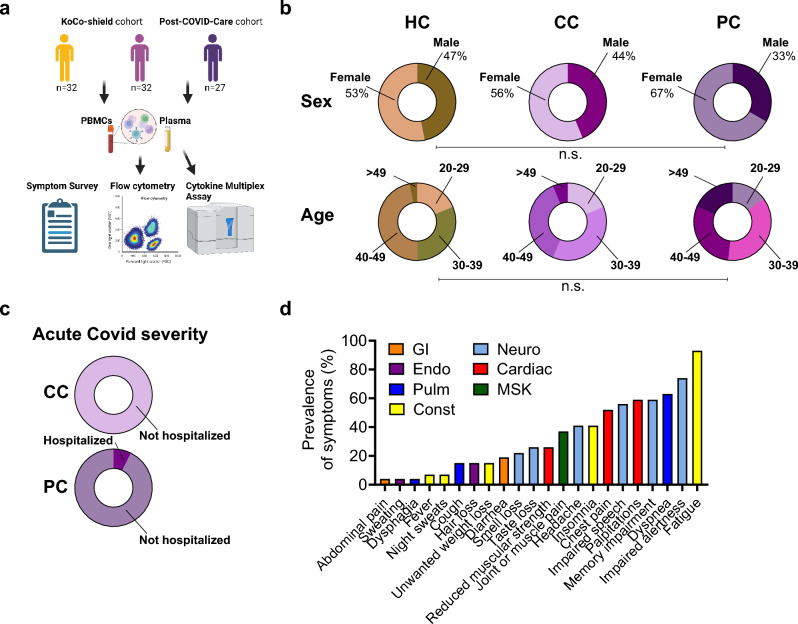
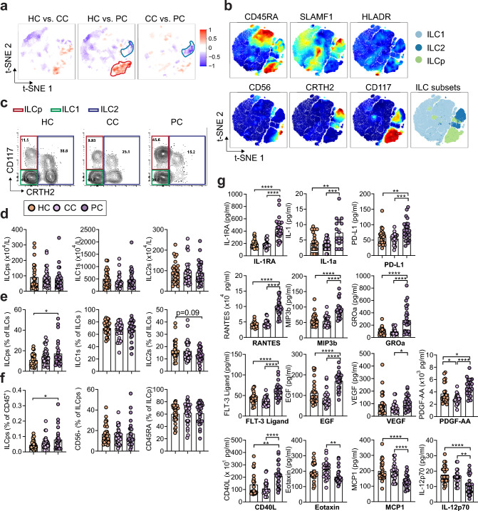
Background: Innate lymphoid cells (ILCs) are key organizers of tissue immune responses and regulate tissue development, repair, and pathology. Persistent clinical sequelae beyond 12 weeks following acute COVID-19 disease, named post-COVID syndrome (PCS), are increasingly recognized in convalescent individuals. ILCs have been associated with the severity of COVID-19 symptoms but their role in the development of PCS remains poorly defined.

Methods and results: Here, we used multiparametric immune phenotyping, finding expanded circulating ILC precursors (ILCPs) and concurrent decreased group 2 innate lymphoid cells (ILC2s) in PCS patients compared to well-matched convalescent control groups at > 3 months after infection or healthy controls. Patients with PCS showed elevated expression of chemokines and cytokines associated with trafficking of immune cells (CCL19/MIP-3b, FLT3-ligand), endothelial inflammation and repair (CXCL1, EGF, RANTES, IL-1RA, PDGF-AA).

Conclusion: These results define immunological parameters associated with PCS and might help find biomarkers and disease-relevant therapeutic strategies.

Keywords: COVID-19; Immune activation; Innate lymphoid cells; Post-COVID-19-syndrome; Tissue immunology.


Conflict of interest statement

The authors declare no commercial or financial conflicts of interest.

Figures

Fig. 1
Fig. 1
Clinical characteristics of study cohorts. a Overview of study cohorts and methods. The figure is partly created with BioRender.com. b Demographic data for healthy, uninfected controls (HC), convalescent SARS-CoV-2 participants without persisting symptoms (CC) and convalescent SARS-CoV-2 participants with persisting symptoms (PC) displayed as ring charts. Statistical significance is shown by capped lines as Chi-square tests for ‘Sex’ and post hoc comparisons for ‘Age’. Further characteristics are detailed in Table 1. c Percentage of hospitalization during acute COVID infection for CC and PC participants displayed as ring charts. d Prevalence of top 22 self-reported symptoms in PC participants (least prevalent (left) to most prevalent (right)). Symptoms are colored according to physiological systems. Gastrointestinal (GI), endocrine (Endo), pulmonary (Pulm), constitutional (Const), neurological (Neuro), cardiac, and musculoskeletal (MSK)
Fig. 2
Fig. 2
Post-COVID participants show altered cytokine expression and levels of innate lymphoid cells. a High-dimensionality reduction analysis of innate lymphoid cells (ILCs, gated as lymphocytes, singlets, and CD45+CD3LinCD127+ cells as shown in Suppl. Fig. 1) from peripheral blood mononuclear cells (PBMCs) of HC, CC, and PC groups. High-resolution group differences were visualized by calculating Cohen's D for a given comparison across the t-SNE map. Residual plot showing differences between maps. Phenotypes within red circles were confirmed to be statistically more common in PC samples, and phenotypes within blue circles were less common in PC samples. Analysis is based on flow cytometry data from 32 HC, 32, CC, and 27 PC samples. b Relative expression intensities (combined HC, CC, and PC samples) of parameters used in the t-SNE analysis. cf Representative flow cytometry plots (c) and quantification (df), showing total numbers (d) and percent (e, f) innate lymphoid cell populations in HC, CC, and PC groups at 3–10 months after acute COVID infection. g Multiplex assay quantification showing plasma levels of IL-1RA, IL-1a, PDL-1, RANTES, MIP-3b, Groa, FLT3 Ligand, EGF, VEGF, PDGF-AA, CD40L, Eotaxin, MCP1, and IL-12p70 in healthy controls with no prior SARS-CoV-2 infection (HC), convalescent SARS-CoV-2 participants without persisting symptoms (CC), and convalescent SARS-CoV-2 participants with persisting symptoms (PC) at 3–10 months after acute COVID infection

References

    1. Clark DV, Kibuuka H, Millard M, Wakabi S, Lukwago L, Taylor A, et al. Long-term sequelae after Ebola virus disease in Bundibugyo, Uganda: a retrospective cohort study. Lancet Infect Dis. 2015;15:905–912. doi: 10.1016/S1473-3099(15)70152-0. - DOI - PubMed
    1. A Post-Graduate Lecture ON THE NERVOUS SEQUELÆ OF INFLUENZA. Lancet 1893;142:73–6. 10.1016/S0140-6736(00)65088-2.
    1. Fluge Ø, Tronstad KJ, Mella O. Pathomechanisms and possible interventions in myalgic encephalomyelitis/chronic fatigue syndrome (ME/CFS). J Clin Invest 2021. 10.1172/JCI150377. - PMC - PubMed
    1. Newton DJ, Kennedy G, Chan KKF, Lang CC, Belch JJF, Khan F. Large and small artery endothelial dysfunction in chronic fatigue syndrome. Int J Cardiol. 2012;154:335–336. doi: 10.1016/j.ijcard.2011.10.030. - DOI - PubMed
    1. Datta SD, Talwar A, Lee JT. A proposed framework and timeline of the spectrum of disease due to SARS-CoV-2 infection: illness beyond acute infection and public health implications. JAMA. 2020;324:2251–2252. doi: 10.1001/jama.2020.22717. - DOI - PubMed

LinkOut - more resources