. 2024 Jun;29(6):1844-1856.
doi: 10.1038/s41380-024-02434-7. Epub 2024 Feb 8.

Cell type-specific expression, regulation and compensation of CDKL5 activity in mouse brain

Affiliations

Cell type-specific expression, regulation and compensation of CDKL5 activity in mouse brain

Margaux Silvestre et al. Mol Psychiatry. 2024 Jun.

Abstract

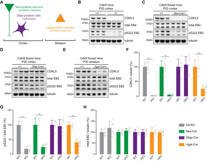
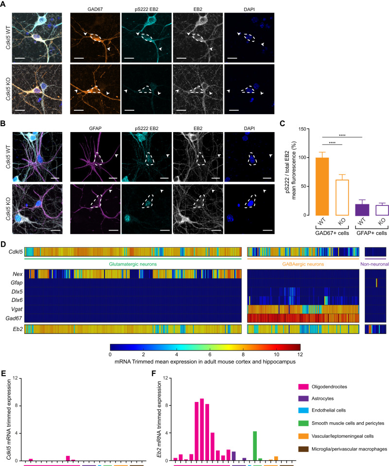
CDKL5 is a brain-enriched serine/threonine kinase, associated with a profound developmental and epileptic encephalopathy called CDKL5 deficiency disorder (CDD). To design targeted therapies for CDD, it is essential to determine where CDKL5 is expressed and is active in the brain and test if compensatory mechanisms exist at cellular level. We generated conditional Cdkl5 knockout mice in excitatory neurons, inhibitory neurons and astrocytes. To assess CDKL5 activity, we utilized a phosphospecific antibody for phosphorylated EB2, a well-known substrate of CDKL5. We found that CDKL5 and EB2 pS222 were prominent in excitatory and inhibitory neurons but were not detected in astrocytes. We observed that approximately 15-20% of EB2 pS222 remained in Cdkl5 knockout brains and primary neurons. Surprisingly, the remaining phosphorylation was modulated by NMDA and PP1/PP2A in neuronal CDKL5 knockout cultures, indicating the presence of a compensating kinase. Using a screen of candidate kinases with highest homology to the CDKL5 kinase domain, we found that CDKL2 and ICK can phosphorylate EB2 S222 in HEK293T cells and in primary neurons. We then generated Cdkl5/Cdkl2 dual knockout mice to directly test if CDKL2 phosphorylates EB2 in vivo and found that CDKL2 phosphorylates CDKL5 substrates in the brain. This study is the first indication that CDKL2 could potentially replace CDKL5 functions in the brain, alluding to novel therapeutic possibilities.


Conflict of interest statement

Part of the findings were included in a patent application by the Francis Crick Institute.

Figures

Fig. 1
Fig. 1. CDKL5 is expressed and active in excitatory and inhibitory neurons of 20-day old mice brains.
A Using a Cre-Lox recombination system, CDKL5 was selectively knocked out in excitatory neurons (Nex-Cre; green), in inhibitory neurons (Vgat-Cre; orange), in astrocytes (Gfap-Cre; purple), or in every cell type (Cdkl5 full KO; gray) and cortex or striatum were collected at P20. BE Western blots showing expression of CDKL5, total EB2 and tubulin, and level of EB2 S222 phosphorylation (pS222) in, respectively, Cdkl5 full KO, Nex-Cre cKO, Gfap-Cre cKO and Vgat-Cre cKO mice at P20. FH Quantification of CDKL5 expression, EB2 pS222 and total EB2 expression in P20 Cdkl5 full KO, Nex-Cre cKO, Gfap-Cre cKO and Vgat-Cre cKO mice. Mann–Whitney test. n = 3 animals per genotype with 2 technical replicates. **p ≤ 0.01. *p ≤ 0.05.
Fig. 2
Fig. 2. EB2 phosphorylation is present in inhibitory neurons but not in astrocytes of mouse primary neurons.
A Cdkl5 KO and WT cortical neurons at DIV8 in culture were co-stained with GAD67, an inhibitory neuron marker, EB2 pS222, total EB2 and DAPI. White arrows indicate neuronal dendrites and dashed lines indicates cell body. Scale bar is 15 μm. B Cdkl5 KO and WT cortical neurons at DIV8 in culture were co-stained with GFAP, an astrocyte marker, EB2 pS222, total EB2 and DAPI. Scale bar is 15 μm. Dashed lines indicates cell body. C Quantification of EB2 phosphorylation mean fluorescence in GAD67- and GFAP-positive cells of Cdkl5 WT and KO cortical neurons. n = 11–16 cells. Mann–Whitney test. ****p ≤ 0.0001. D Expression of Cdkl5 and different cell markers in mouse adult isocortex and hippocampus from single-cell RNA sequencing data. E, F Closer view of the expression of Cdkl5 and Eb2 respectively in different non-neuronal cells of adult mouse isocortex and hippocampus from single-cell RNA sequencing data.
Fig. 3
Fig. 3. EB2 S222 phosphorylation is reduced by synaptic NMDAR-dependent calcium influx.
A Quantification of EB2 pS222 levels upon 50 μM NMDA treatment of DIV14 rat primary cortical neurons. Welch’s t test. n = 3 technical replicates. B Quantification of EB2 pS222 levels upon 10 μM glycine and 25 μM glutamate treatment of DIV15 rat primary cortical neurons. Welch’s t test. n = 4 technical replicates. C Quantification of EB2 pS222 levels upon 50 μM NMDA treatment of DIV15 rat primary cortical neurons in normal ACSF and in calcium free ACSF. Dunnett’s multiple comparisons test. n = 4 technical replicates. D Quantification of EB2 pS222 levels upon 10 μM MK801 and 50 μM bicuculline pre-treatment (10 min) and 50 μM NMDA treatment of DIV15 rat primary cortical neurons. Dunnett’s multiple comparisons test. n = 4 technical replicates.
Fig. 4
Fig. 4. Inhibition of PP1 and PP2A increases EB2 phosphorylation in presence and absence of CDKL5.
A Western blots showing CDKL5, total EB2 and tubulin expression and EB2 pS222 level upon 1 μM okadaic acid (OA) for 40 min, 20 μM cyclosporin A (CSA) for 40 min and 50 μM NMDA treatment for 20 min in DIV14-15 rat primary cortical culture. B Quantification of EB2 pS222 level upon 1 μM OA for 40 min, 20 μM CSA for 40 min and 50 μM NMDA treatment for 20 min in DIV14-15 rat cortical primary culture. Mann–Whitney test. n = 5 with 2 technical replicates per treatment. ****p < 0.0001. **p ≤ 0.01. *p ≤ 0.05. C Western blots showing total EB2 and tubulin expression and EB2 pS222 level upon 1 μM OA treatment for 40 min, 50 μM NMDA treatment for 20 min and a combination of OA and NMDA treatments (1 μM OA treatment for 20 min followed by 20 min of OA + NMDA) in DIV8 mouse Cdkl5 WT and KO primary cortical culture. D Quantification of EB2 S222 phosphorylation level upon 1 μM OA treatment for 40 min, 50 μM NMDA treatment for 20 min and a combination of OA and NMDA treatments in DIV8 mouse Cdkl5 WT and KO primary cortical culture. Mann–Whitney test. n = 3 with 2 technical replicates per treatment. **p ≤ 0.01. *p ≤ 0.05. Only significance within WT treatments and within KO treatments are shown. E Pairwise quantification of EB2 S222 phosphorylation upon 1 μM OA treatment for 40 min in Cdkl5 KO primary cortical culture. Mann–Whitney test. n = 3 with 2 technical replicates per treatment. *p ≤ 0.05.
Fig. 5
Fig. 5. EB2 phosphorylation by ICK and CDKL2 in HEK293T cells and primary neurons.
A CDKL5 and its closely-related kinases on the human phylogenetic tree. B Western blot showing levels of FLAG, total EB2 and EB2 pS222 in HEK293T cells co-transfected with StrepII-tagged EB2 and FLAG-tagged ICK WT, MAK WT, MOK WT, CDKL1 WT, CDKL2 WT, CDKL3 WT, CDKL4 WT, CDKL5 WT or CDKL5 kinases-dead (KD). C Western blot showing levels of FLAG, total EB2 and EB2 pS222 in HEK293T cells co-transfected with StrepII-tagged EB2 and FLAG-tagged ICK WT/KD, CDKL2 WT/KD or CDKL5 WT/KD. DF EB2 pS222 quantification comparing WT and KD of ICK, CDKL2 and CDKL5 expressing cells respectively. n = 6 per condition. Mann–Whitney test. **p ≤ 0.01. G DIV8 Cdkl5 KO primary mouse hippocampal neurons, overexpressing FLAG-tagged ICK, CDKL2 or CDKL5 WT, were co-stained with FLAG, EB2 pS222, total EB2 and DAPI. Full lines highlight neurons expressing the kinases and dashed lines highlight neurons which do not express the kinases. Scale bar is 30 μm. H Quantification of EB2 pS222 mean fluorescence in Cdkl5 WT neurons, in Cdkl5 KO neurons and in Cdkl5 KO neurons overexpressing the three kinases. n = 21–166 per condition. Mann–Whitney test. ****p ≤ 0.0001. I Quantification of total EB2 mean fluorescence in Cdkl5 WT neurons, in Cdkl5 KO neurons and in Cdkl5 KO neurons overexpressing the three kinases. n = 21–166 per condition. Mann–Whitney test. ****p ≤ 0.0001.
Fig. 6
Fig. 6. CDKL2 phosphorylates CDKL5 substrates in mouse brain.
A Schematic of the generation of the dual Cdkl5/Cdkl2 KO mouse model. An insert of 700 bp containing the exon 4 of Cdkl2 was deleted from Cdkl5 KO embryos using CRISPR-Cas9. B Western blot showing levels of CDKL5, total EB2 pS222, tubulin and CDKL2 in the cortex of P10 dual Cdkl5/Cdkl2 KO animals. The arrow indicates CDKL2, [*] indicates a non-specific band. C Quantification of EB2 pS222 levels in the cortex of P10 Cdkl5/Cdkl2 KO animals. n = 3 animals per genotype with 2 technical replicates. Mann–Whitney test. **p ≤ 0.01. D Western blot showing levels of total MAP1S and MAP1S pS812 in the cortex of P10 dual Cdkl5/Cdkl2 KO animals. E Schematic encapsulating our findings. CDKL5 and CDKL2 phosphorylate EB2 on residue S222, a process modulated by calcium entry through NMDA receptors. Additionally, the dephosphorylation of EB2 S222 is attributed to the phosphatases PP1 and PP2A.

References

    1. Manning G, Whyte DB, Martinez R, Hunter T, Sudarsanam S. The protein kinase complement of the human genome. Science. 2002;298:1912–34. 10.1126/science.1075762 - DOI - PubMed
    1. Leonard H, Downs J, Benke TA, Swanson L, Olson H, Demarest S. CDKL5 deficiency disorder: clinical features, diagnosis, and management. Lancet Neurol. 2022;21:563–76. 10.1016/S1474-4422(22)00035-7 - DOI - PMC - PubMed
    1. Olson HE, Demarest ST, Pestana-Knight EM, Swanson LC, Iqbal S, Lal D, et al. Cyclin-dependent kinase-like 5 deficiency disorder: clinical review. Pediatr Neurol. 2019;97:18–25. 10.1016/j.pediatrneurol.2019.02.015 - DOI - PMC - PubMed
    1. Scala E, Ariani F, Mari F, Caselli R, Pescucci C, Longo I, et al. CDKL5/STK9 is mutated in Rett syndrome variant with infantile spasms. J Med Genet. 2005;42:103–7. 10.1136/jmg.2004.026237 - DOI - PMC - PubMed
    1. Tao J, Van Esch H, Hagedorn-Greiwe M, Hoffmann K, Moser B, Raynaud M, et al. Mutations in the X-linked cyclin-dependent kinase-like 5 (CDKL5/STK9) gene are associated with severe neurodevelopmental retardation. Am J Hum Genet. 2004;75:1149–54. 10.1086/426460 - DOI - PMC - PubMed

Substances

Supplementary concepts