. 2024 Apr;396(1):57-69.
doi: 10.1007/s00441-024-03864-z. Epub 2024 Feb 8.

Electron microscopic analysis of the influence of iPSC-derived motor neurons on bioengineered human skeletal muscle tissues

Affiliations

Electron microscopic analysis of the influence of iPSC-derived motor neurons on bioengineered human skeletal muscle tissues

Christine T Nguyen et al. Cell Tissue Res. 2024 Apr.

Abstract

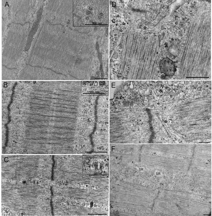
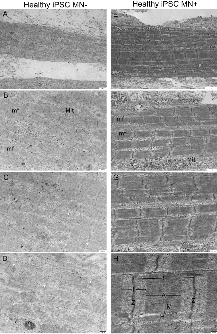
3D bioengineered skeletal muscle macrotissues are increasingly important for studies of cell biology and development of therapeutics. Tissues derived from immortalized cells obtained from patient samples, or from pluripotent stem cells, can be co-cultured with motor-neurons to create models of human neuromuscular junctions in culture. In this study, we present foundational work on 3D cultured muscle ultrastructure, with and without motor neurons, which is enabled by the development of a new co-culture platform. Our results show that tissues from Duchenne muscular dystrophy patients are poorly organized compared to tissues grown from healthy donor and that the presence of motor neurons invariably improves sarcomere organization. Electron micrographs show that in the presence of motor neurons, filament directionality, banding patterns, z-disc continuity, and the appearance of presumptive SSR and T-tubule profiles all improve in healthy, DMD-, and iPSC-derived muscle tissue. Further work to identify the underlying defects of DMD tissue disorganization and the mechanisms by which motor neurons support muscle are likely to yield potential new therapeutic approaches for treating patients suffering from Duchenne muscular dystrophy.

Keywords: 3D culture; Duchenne muscular dystrophy; Muscle; Ultrastructure.


Conflict of interest statement

The authors declare no competing interests.

Figures

Fig. 1
Fig. 1
MyoFMS device fabrication and skeletal muscle tissue production. A 3D model of a 12-well footprint MyoFMS culture plate together with a B 3D model zoom-in and a C CAD drawing of a single well to visualize multi-post arrangement and tissue seeding bed dimensions and orientation. D Representative images of each step in the fabrication process used to generate the final PDMS-based MyoFMS culture device. E (bottom) Timeline showing the steps employed to generate skeletal muscle tissues in MyoFMS. E (left) Representative, 4× stitched confocal image of iPSC-derived skeletal muscle tissue at day 10 of differentiation. Magenta, sarcomeric α-actinin (SAA). Scale bar, 25 mm. E (right) 40× confocal split-channel images of a representative region of the E (left) engineered muscle tissue. Magenta, SAA; cyan, phalloidin; yellow, Draq5. Scale bar, 100 µm
Fig. 2
Fig. 2
Representative comparison electron micrographs of 3D bioengineered macrotissues ± iPSC-derived motor neurons at day 10. AD Healthy AB1167 muscle-alone (n = 3), EH AB1167 co-cultured with iPSC-derived motor neurons (n = 2). A, E Single muscle cell showing multiple myofibrils (mf). Ultrastructural features in both conditions include mitochondria (Mit), sarcomeres (S), A-bands (A), I-bands (I), H-zones (H), M-lines (M), and Z-discs (Z). AD Macrotissues show distinct myofibrils with clear myofilament organization. EH Neuro-muscular macrotissues show slight improvement in sarcomere organization with clearer M-lines and strong crosslinking compared to muscle-alone tissues. Sarcoplasmic reticulum (SR) becomes more prevalent in neuro-muscular macrotissues (white arrow)
Fig. 3
Fig. 3
Ultrastructural features of 3D bioengineered neuromuscular macrotissues at day 10. A, B Healthy Ab1167 co-culture illustrating presumptive skeletal muscle triad with sarcoplasmic reticulum (SR) and t-tubule (T) between myofibril. Scale 500 nm. C Healthy iPSC co-culture illustrating presumptive skeletal muscle triad with sarcoplasmic reticulum (SR) and t-tubule (T) between A- and I-bands. Scale 500 nm. D, E Healthy Ab1167 co-culture illustrating t-tubule invagination through myofibril (asterisk). Scale 500 nm. F DMD 1071 co-culture showing sarcoplasmic reticulum (SR). Scale 200 nm
Fig. 4
Fig. 4
Representative comparison electron micrographs of 3D bioengineered DMD-1071 macrotissues ± iPSC-derived motor neurons at day 10. AD DMD 1071 muscle-alone (n = 3), EH DMD 1071 co-cultured with iPSC-derived motor neurons (n = 4). A, E Single muscle cell. AE DMD 1071 muscle-alone electron micrographs showing the presence of disorganized myofilaments (black arrows). D Myofilaments run in non-uniform, unparalleled directions (yellow dashed lines). Faint and short z-bodies are seen throughout tissues with ill-defined sarcomere boundaries (white arrows). EH DMD 1071 neuro-muscular macrotissues show a significant improvement in sarcomere organization showing the presence of sarcomeres (S), A-bands (A), I-bands (I), and Z-discs (Z), and some faint M-lines (white arrowhead), when compared to muscle-alone
Fig. 5
Fig. 5
Representative comparison electron micrographs of 3D bioengineered macrotissues ± iPSC-derived motor neurons at day 8, or 10. AD Healthy iPSC-derived muscle-alone (n = 3), EH Healthy iPSC-derived muscle co-cultured with iPSC-derived motor neurons (= 3). A, E Single muscle cell showing multiple myofibrils (mf). EH Mitochondria (Mit) is seen in both conditions, whereas faint short Z-disc (Z), less distinct sarcomere (S), and structural feature A-bands (A), I-bands (I), H-zones (H), and M-lines (M) are seen in muscle-alone tissues (AD) compared to neuro-muscular tissues (EH). In the presence of motor neurons, myofilament organization improves, illustrated with clear Z-disc, A–I banding, strong crosslinking with well-defined M-line, and H-zones
Fig. 6
Fig. 6
Representative electron micrographs of presumptive skeletal muscle triads. AC Triad structures are indicated with white arrowheads, whereas mitochondria are false colored in magenta. A, B Muscle-alone iPSC-derived human macro tissues day 10 of differentiation. Scale bar 500 nm. C Healthy Ab1167 co-cultured with iPSC-derived motor neurons day 10 of differentiation. Scale bar 500 nm

References

    1. Bakooshli MA, Lippmann ES, Mulcahy B, Iyer N, Nguyen CT, Tung K, Stewart BA, van den Dorpel H, Fuehrmann T, Shoichet M, Bigot A, Pegoraro E, Ahn H, Ginsberg H, Zhen M, Ashton RS, Gilbert PM. A 3D culture model of innervated human skeletal muscle enables studies of the adult neuromuscular junction. ELife. 2019;8:e44530. doi: 10.7554/eLife.44530. - DOI - PMC - PubMed
    1. Boncompagni S, d'Amelio L, Fulle S, Fanò G, Protasico F. Progressive disorganization of the excitation-contraction coupling apparatus in aging human skeletal muscle as revealed by electron microscopy: a possible role in the decline of muscle performance. J Gerontol A Biol Sci Med Sci. 2006;61(10):995–1008. doi: 10.1093/gerona/61.10.995. - DOI - PubMed
    1. Boncompagni S, Rossi AE, Micaroni M, Beznoussenko GV, Polishchuk RS, Dirksen RT, Protasico F. Mitochondria are linked to calcium stores in striated muscle by developmentally regulated tethering structures. Mol Biol Cell. 2009;20(3):1058–1067. doi: 10.1091/mbc.E08-07-0783. - DOI - PMC - PubMed
    1. Burattini S, Ferri P, Battistelli M, Curci R, Luchetti F, Falcieri E. C2C12 murine myoblasts as a model of skeletal muscle development: morpho-functional characterization. Eur J Histochem. 2004;48(3):223–233. - PubMed
    1. Dessauge F, Schleder C, Perruchot MH, Rouger K. 3D in vitro models of skeletal muscle: myopshere, myobundle and bioprinted muscle construct. Vet Res. 2021;52:72. doi: 10.1186/s13567-021-00942-w. - DOI - PMC - PubMed

LinkOut - more resources