Review
. 2024 Jan;26(1):26-40.
doi: 10.5853/jos.2023.02481. Epub 2024 Jan 30.

Emerging Concept of Intracranial Arterial Diseases: The Role of High Resolution Vessel Wall MRI

Affiliations
Review

Emerging Concept of Intracranial Arterial Diseases: The Role of High Resolution Vessel Wall MRI

Dong-Wan Kang et al. J Stroke. 2024 Jan.

Abstract

Intracranial arterial disease (ICAD) is a heterogeneous condition characterized by distinct pathologies, including atherosclerosis. Advances in magnetic resonance technology have enabled the visualization of intracranial arteries using high-resolution vessel wall imaging (HR-VWI). This review summarizes the anatomical, embryological, and histological differences between the intracranial and extracranial arteries. Next, we review the heterogeneous pathophysiology of ICAD, including atherosclerosis, moyamoya or RNF213 spectrum disease, intracranial dissection, and vasculitis. We also discuss how advances in HR-VWI can be used to differentiate ICAD etiologies. We emphasize that one should consider clinical presentation and timing of imaging in the absence of pathology-radiology correlation data. Future research should focus on understanding the temporal profile of HR-VWI findings and developing quantitative interpretative approaches to improve the decision-making and management of ICAD.

Keywords: Arterial dissection; Atherosclerotic plaque; Intracranial arterial diseases; Magnetic resonance imaging; Moyamoya disease.


Conflict of interest statement

Conflicts of interest

The authors have no financial conflicts of interest.

Figures
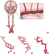
Figure 1.
Figure 1.
Overview of cerebrovascular anatomy and embryology. (A) Schematic drawing of the anatomy of the intracranial and extracranial arteries. The middle cerebral artery traverses through the subarachnoid space and extends perforators into the cerebral parenchyma. (B) In the early embryo, the three aortic arches (AA) link the ventral (VA) and dorsal aorta (DA). (C) As the first (I AA) and second aortic arches (II AA) regress, the DA and the third aortic arch (III AA) remain as the primitive internal carotid artery (ICA). The remnant I AA corresponds to the mandibular artery (MA), and the remnant II AA corresponds to the hyoid artery (HA). Other embryonic arteries arise, such as the primitive maxillary artery (PMA), trigeminal artery (TA), and primitive dorsal ophthalmic artery (PDOA). (D) The embryonic arteries that regress divide the primitive ICA into seven segments. The first segment corresponds to the remnant of the III AA. The second segment is the remnant of the DA between the II AA and III AA. The third to seventh segments are divided into the MA, PMA, PDOA, primitive ventral OA (PVOA), and ICA bifurcation. CrD, cranial division; CaD, caudal division; HypA, hypoglossal artery; PA, proatlantal artery; DAr, dorsal aorta right; DAl, dorsal aorta left; BA, basilar artery; ACA, anterior cerebral artery; PComA, posterior communicating artery; ECA, external carotid artery.
Figure 2.
Figure 2.
Schematic diagram displaying the heterogeneity of intracranial arterial diseases. (A) The structure of a normal intracranial vessel wall consists of endothelial cells, intima, internal elastic lamina (IEL), media, and adventitia. There is no external elastic lamina in intracranial arteries. Intracranial arteries have fewer elastin fibers in the media, thinner adventitia, and thicker IEL compared to extracranial arteries. (B) Atherosclerosis is characterized by a lipid core covered by fibrous caps. Infiltrating macrophages are found within the lipid core. More advanced forms of atherosclerosis are infrequently seen in intracranial arteries. (C) Intracranial arteries in moyamoya disease undergo negative remodeling due to fibrosis and thickening of the intima and proliferation of smooth muscle cells. There is also infolding and chronic contraction of the IEL. (D) Intracranial arterial dissection. The intima is torn inward to form an intimal flap and a double lumen. Intramural hematoma is sometimes seen. (E) Mononuclear and granulomatous adventitial inflammation and focal fibrin thrombus formation in vasculitis. Concentric thickening of the vessel wall is seen. (F) In reversible cerebral vasoconstriction syndrome, the diameter of the entire vessel wall is reduced due to the contraction of smooth muscle in the media layer.
Figure 3.
Figure 3.
A representative image of high-resolution vessel wall imaging of normal intracranial arteries. (A) Time-of-flight image. (B-D) Proton density (PD), T1-weighted, and T1-weighted with gadolinium (T1GD) coronal images showing the left middle cerebral artery (MCA). (E-G) PD, T1-weighted, and T1GD coronal image showing a cross-sectional view of the left MCA.
Figure 4.
Figure 4.
Representative high-resolution vessel wall imaging (HR-VWI) images of various intracranial arterial diseases. (A) HR-VWI findings of atherosclerosis in a 45-year-old male with prehypertension and hypercholesterolemia. A stenotic portion (arrow) at the left middle cerebral artery (MCA) is identified on the time-of-flight (TOF) image. The proton density (PD) image shows no total vessel wall diameter stenosis. The T1 image shows eccentric vessel wall thickening with isointense signal suggesting a lipid core or fibrous cap. The area appears to be enhanced on the T1-weighted with gadolinium (T1GD) image. (B) HR-VWI in a 35-year-old female with moyamoya disease presenting with right MCA infarction. A heterozygous RNF213 c.14429G>A mutation was found. The TOF image shows stenosis at both distal internal carotid artery (ICA) to proximal A1 and M1. The arrow indicates the most stenotic part of the right MCA. Concentric wall thickening involving the right MCA with enhancement and negative remodeling was noted on PD, T1, and T1GD images. (C) HR-VWI of a left MCA dissection in a 48-year-old male who underwent endovascular treatment due to a left MCA infarction 4 months earlier. TOF-magnetic resonance angiography (MRA) shows a stenotic portion of the left MCA (arrow). The intimal flap is clearly visible on the PD axial image. Angiography at the time of endovascular treatment shows the intimal flap (arrow). PD, T1, and T1GD sagittal images show a double lumen and an intramural hematoma. A lesion with hyperintense PD and hyperintense T1 signal and no enhancement suggests a late subacute stage of the intramural hematoma. (D) HR-VWI in a 47-year-old female with vasculitis. TOF image shows a severely stenotic portion of the left MCA (arrow). PD, T1, and T1GD coronal images show wall thickening and prominent enhancement of the left M1. Sagittal images show concentric wall thickening with negative remodeling with strong enhancement. The stenosis later progressed despite steroid treatment. (E) HR-VWI in a 27-year-old female with reversible cerebral vasoconstriction syndrome presenting with severe headache. TOF-MRA axial image shows multifocal stenoses of both MCAs. Reconstructed TOF-MRA shows multifocal mild to moderate stenoses at the distal ICA, MCA, anterior cerebral artery, and basilar artery. Sagittal images of the right MCA show normal appearance of the vessel wall. Coronal images show stenotic segments without any abnormal vessel wall signal. Intracranial vessels were significantly improved on follow-up HR-VWI.

References

    1. Gorelick PB, Wong KS, Bae HJ, Pandey DK. Large artery intracranial occlusive disease: a large worldwide burden but a relatively neglected frontier. Stroke. 2008;39:2396–2399. - PubMed
    1. Derdeyn CP, Chimowitz MI, Lynn MJ, Fiorella D, Turan TN, Janis LS, et al. Aggressive medical treatment with or without stenting in high-risk patients with intracranial artery stenosis (SAMMPRIS): the final results of a randomised trial. Lancet. 2014;383:333–341. - PMC - PubMed
    1. Flusty B, de Havenon A, Prabhakaran S, Liebeskind DS, Yaghi S. Intracranial atherosclerosis treatment: past, present, and future. Stroke. 2020;51:e49–e53. - PMC - PubMed
    1. Bala F, Cimflova P, Singh N, Zhang J, Kappelhof M, Kim BJ, et al. Impact of vessel tortuosity and radiological thrombus characteristics on the choice of first-line thrombectomy strategy: results from the ESCAPE-NA1 trial. Eur Stroke J. 2023;8:675–683. - PMC - PubMed
    1. Bang OY, Toyoda K, Arenillas JF, Liu L, Kim JS. Intracranial large artery disease of non-atherosclerotic origin: recent progress and clinical implications. J Stroke. 2018;20:208–217. - PMC - PubMed

LinkOut - more resources