. 2024 Feb 8;43(1):45.
doi: 10.1186/s13046-023-02929-1.

RNA-binding protein RPS7 promotes hepatocellular carcinoma progression via LOXL2-dependent activation of ITGB1/FAK/SRC signaling

Affiliations

RNA-binding protein RPS7 promotes hepatocellular carcinoma progression via LOXL2-dependent activation of ITGB1/FAK/SRC signaling

Yu-Jiao Zhou et al. J Exp Clin Cancer Res. .

Erratum in

Abstract

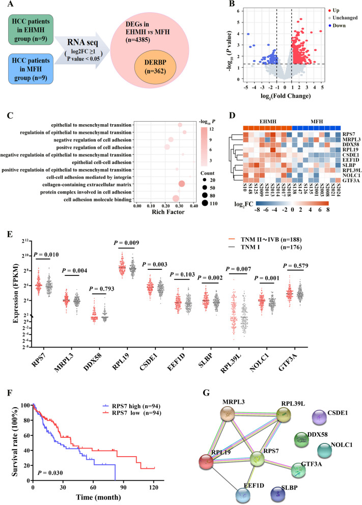
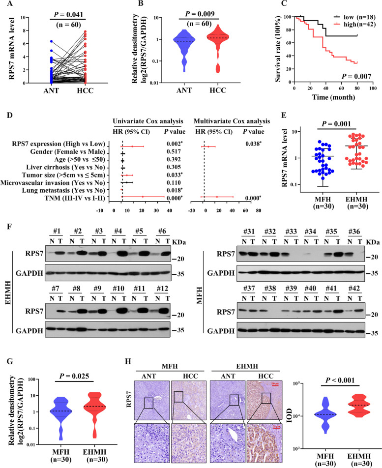
Background: Metastasis is one of the leading cause contributes to treatment failure and poor prognosis of hepatocellular carcinoma (HCC) patients. The underlying mechanism of HCC metastasis remains to be determined. Although several RNA binding proteins (RBPs) have been found to participate in tumorigenesis and progression of liver cancer, the role of RBPs in HCC patients with extrahepatic metastases is poorly understood.

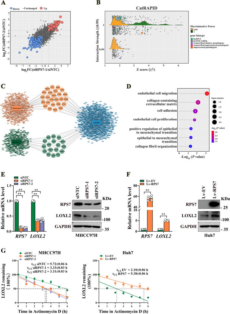
Methods: By performing RNA-seq of primary HCC tissues (including HCC with extrahepatic metastasis and those did not develop metastasis), we identified a set of HCC metastasis-associated RBPs candidates. Among which, ribosomal protein S7 (RPS7) was found to be remarkably increased in HCC tissues and be strongly related to HCC poor survival. Overexpression or CRISPR-Cas9-mediated knockout were applied to investigate the role of RPS7 on the metastasis-associated phenotypes of HCC cells. RNA sequencing, RIP, RNA-pull down, dual luciferase reporter assay, nascent RNA capture assay, and RNA decay and so on, were applied to reveal the underlying mechanism of RPS7 induced HCC metastasis.

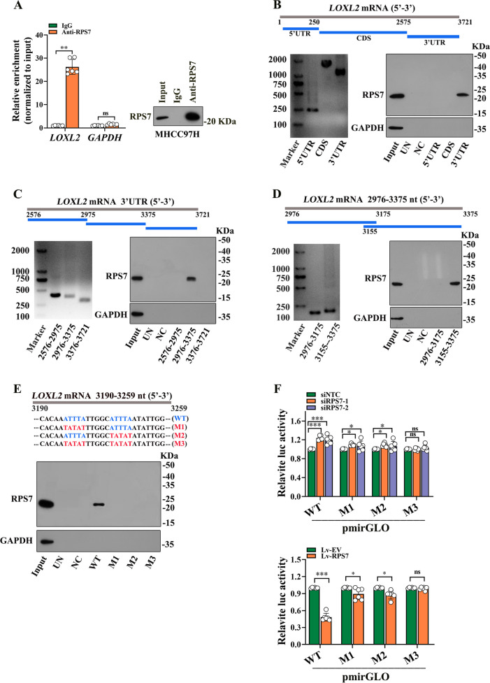
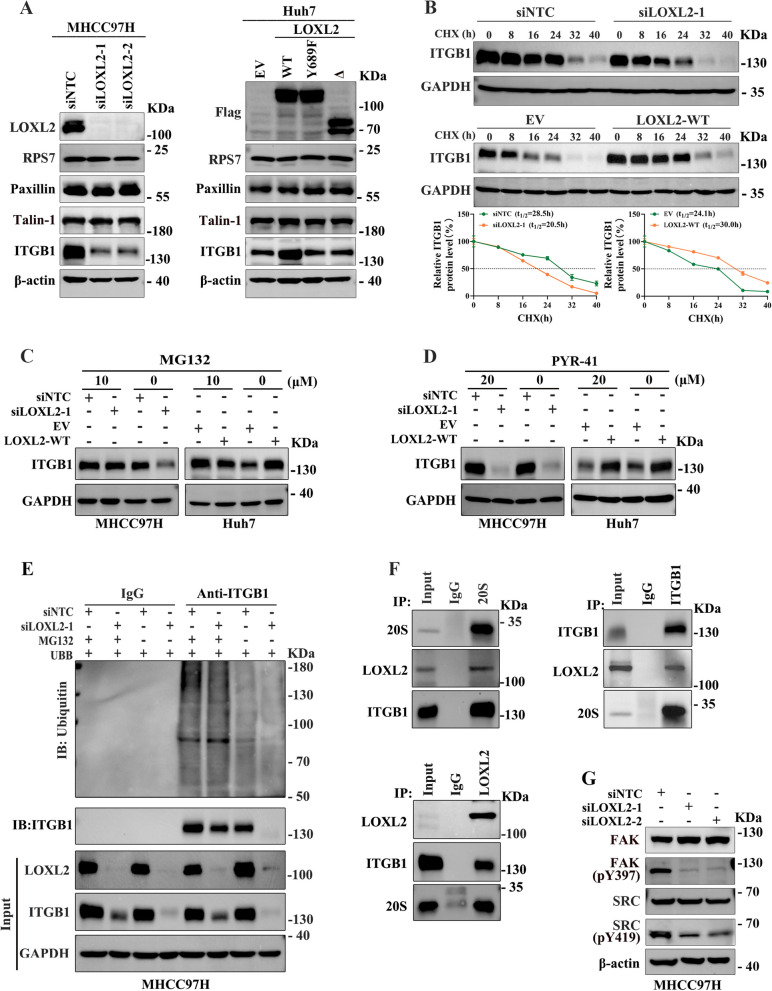
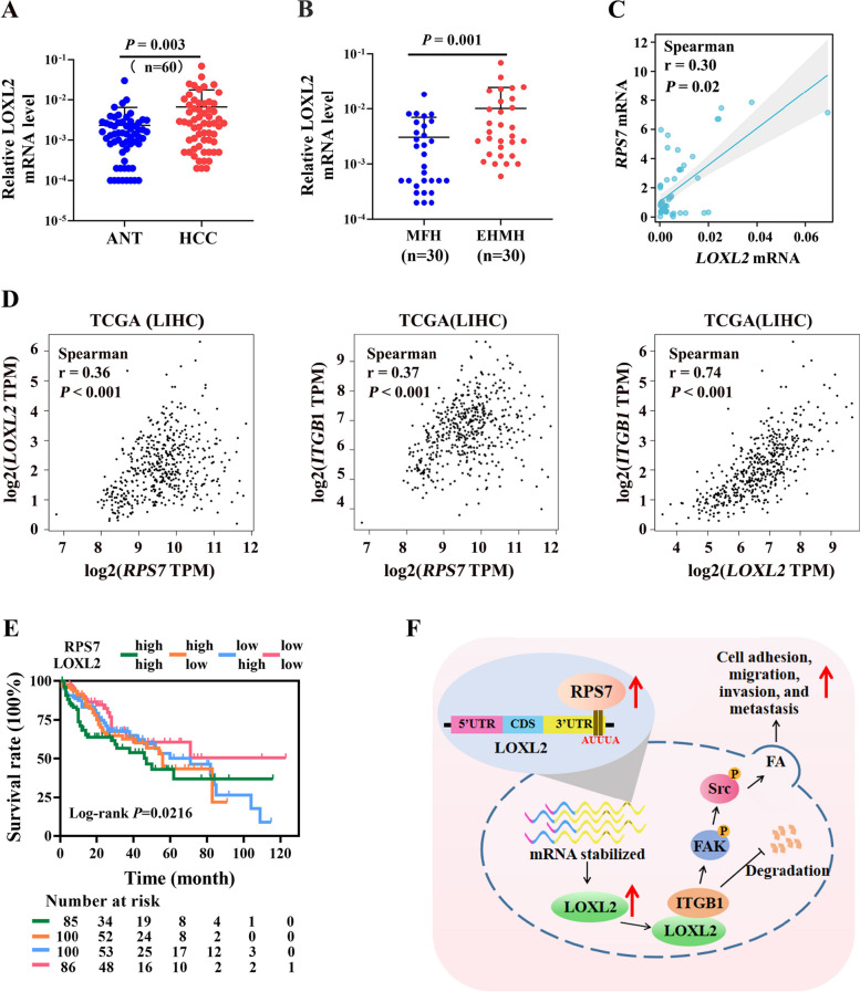
Results: Gain- and loss- of function analyses revealed that RPS7 promoted HCC cells adhesion, migration and invasion capabilities, as well as lung metastasis. Mechanistically, we uncovered that lysyl oxidase-like 2 (LOXL2) was a critical downstream target of RPS7. RPS7 could stabilize LOXL2 mRNA by binding to AUUUA motifs in the 3155-3375 region of the 3'UTR of LOXL2 mRNA, thus increased LOXL2 expression via elevating LOXL2 mRNA abundance. Further research revealed that LOXL2 could accelerate focal adhesion formation through maintaining the protein stability of ITGB1 and activating ITGB1-mediated FAK/SRC signaling pathway, and thereby contribute to the pro-metastasis effect of RPS7.

Conclusions: Taken together, our data reveal a novel function of RPS7 in HCC metastasis, also reveal the critical roles of the RPS7/LOXL2/ITGB1 axis in HCC metastasis and shed new light on the exploration of molecular drugs against HCC.

Keywords: Focal adhesion; Hepatocellular carcinoma; Lysyl oxidase-like 2; Metastasis; RNA-binding protein; Ribosomal protein S7.


Conflict of interest statement

The authors have no conflicts of interest to declare.

Figures

Fig. 1
Fig. 1
Identification of RPS7 as an important RNA-binding protein (RBP) that associates with HCC progression. Nine pairs of primary HCC tissues with extrahepatic metastasis (EHMH) and nine pairs of metastasis-free HCC tissues (MFH) as well as corresponding adjacent nontumoral tissues (ANT) were used for RNA sequencing analysis, aiming to screen and identify RBP that significantly correlate with HCC metastasis. The clinical data of HCC patients from TCGA-LIHC datasets were used for validation. According to the tumor-node-metastasis (TNM) staging system for liver cancer, patients were classified into metastasis group (TNM II~IVB) and metastasis-free group (TNM I ). A Schematic screening workflow of differentially expressed RBPs (DERBPs) in EHMH compared to MFH. B Volcano plot for DERBPs expression. C Go analysis of 312 upregulated DERBPs. D Heat map for the top 10 upregulated DERBPs. E The expression difference of each gene was analyzed by comparing metastasis group with metastasis-free group based on TCGA database. F The correlation between RPS7 levels and overall survival rate in HCC with metastasis. G The PPI network by String database
Fig. 2
Fig. 2
RPS7 is up-regulated in HCC with metastasis and is correlated with poor survival. Sixty pairs of HCC samples and matched adjacent nontumoral tissues (ANT) were used to detect the expression of RPS7. Among which, thirty pairs were tissues came from HCC patients with extrahepatic metastasis and other 30 pairs were from metastasis-free HCC patients, designated as EHMH and MFH, respectively. A The mRNA levels of RPS7 in HCC tissues compared to matched ANT. B The protein levels of RPS7 in HCC tissues compared to matched ANT. C The correlation between RPS7 levels and HCC overall survival was evaluated by KM curve. D Forest plot represented the correlation between RPS7 expression and HCC survival, which was evaluated by using univariate and multivariate Cox regression analyses. *, P < 0.05. E The comparison of RPS7 mRNA levels between EHMH group and MFH group. F RPS7 protein levels in EHMH and MFH were respectively detected by western blot assay, as shown was the representative result. G The comparison of RPS7 protein levels between EHMH group and MFH group. H. The expression and localization of RPS7 in HCC tissues compared to matched ANT were analyzed by Immunohistochemistry
Fig. 3
Fig. 3
RPS7 knockout inhibits HCC cell adhesion, migration and invasion in vitro and metastasis in vivo. MHCC97H and HLE cells, two highly invasive HCC cell lines were used to establish stable RPS7 knockout cells by using CRISPR/Cas9 system (RPS7 KO). Non-target knockout cells (NTC KO) were correspondingly used as controls. The cell-matrix adhesion capacity, migration and invasion ability of cells in vitro as well as metastasis in vivo were observed. A The knockout efficiency was determined by western blot assay. B RPS7-knockout MHCC97H and HLE cells adhesion to fibronectin, collagen I and collagen IV were detected using cell-matrix adhesion assay. C The effect of RPS7-knockut on cell migration and invasion were determined by Transwell assay. D Orthotopic mouse models were constructed using RPS7-knockout MHCC97H cells and control cells (each group, n=12). The effect of RPS7-knockout on tumor size and numbers were evaluated. E and F The effects of RPS7-knockout on lung metastasis were evaluated by orthotopic mouse models and tail vein lung metastasis mouse models, respectively. Representative data are from at least 3 independent experiments. Data are shown as mean ± SD. **, P < 0.01
Fig. 4
Fig. 4
LOXL2, a key downstream target of RPS7, is regulated by RPS7 at post-transcriptional level. To elucidate the potential mechanism underlying RPS7-mediated HCC metastasis, RAN-seq analysis was performed using two independent RPS7-knockdown (siRPS7-1 and siRPS7-2) and non-target knockdown (siNTC) MHCC97H cells. A As shown were differentially expressed genes after RPS7 knockdown. B Candidate transcripts that may bind to RPS7 protein were extracted from catRAPID database. C After combing the RNA-seq data and catRAPID data set, differentially expressed genes that possibly bind to RPS7 protein were identified. D KEGG analysis of the candidate downstream genes. E RPS7 knockdown decreased the mRNA and protein levels of LOXL2. F. RPS7 overexpression increased the mRNA and protein levels of LOXL2. G. The effect of RPS7 on LOXL2 mRNA half-life was determined by RNA decay assay. **, P < 0.01
Fig. 5
Fig. 5
RPS7 regulates LOXL2 expression via binding to LOXL2 mRNA. A RNA immunoprecipitation assay was performed in MHCC97H cells by using antibody targeting RPS7, qRT-PCR was performed to detect the mRNA level of LOXL2 in the precipitated RNA-Protein complex. B-E To further identify the specific binding region of RPS7 on LOXL2 mRNA, a series of RNA pull-down analyses were performed using different biotinylated fragments of LOXL2 mRNA including 5’UTR, CDS, 3’UTR (B), and 2576-2975 nt, 2976-3375 nt, 3376-3721 nt (C), 2976-3175 nt, 3155-3375 nt (D) within 3’UTR, as well as truncated recombinants of 3190-3259 nt (E). Western blot assay was subsequently used to detect RPS7 in these indicated mRNA fragments pull-down complex. Input showed 1% lysate. UN represented a control using beads only. NC represented a negative control using non-blot RNA. F. The pmirGLO-derived luciferase reporter containing wild-type and three different mutants of AUUUA motifs were constructed, 293T cells were transfected with these plasmids respectively. Luciferase activities normalized against Renilla luciferase activities were measured to determine the binding efficient of the AUUUA motifs to RPS7 in response to RPS7 overexpression or RPS7 knockdown. *, P < 0.05, **, P < 0.01, ***, P < 0.001. ns, no significant
Fig. 6
Fig. 6
LOXL2 promotes focal adhesion (FA) formation, migration and invasion of HCC cells. A and B To evaluate the role of LOXL2 in HCC metastasis, two pcDNA3.1-LOXL2-3×Flag mutant plasmids, catalytic activity deletion mutant of LOXL2 (LOXL2-Δ) and catalytically inactive point mutant of LOXL2 (LOXL2-Y689F), as well as wild type LOXL2 (LOXL2-WT) were respectively constructed and stably transfected into Huh7 cells. The effect of LOXL2 overexpression on cell-matrix adhesion ability (A) and migration and invasion (B) were analyzed. C The effect of LOXL2 silencing on cell-matrix adhesion ability in MHCC97H cells. D The effect of LOXL2 silencing on cell migration and invasion in MHCC97H cells. E-G. The effect of LOXL2 silencing (E), 20 μM CMMH (F), and LOXL2 overexpression (G) on FA formation in HCC cells were respectively detected by Immunofluorescence staining experiments. Representative data are from at least 3 independent experiments. Data are shown as mean ± SD. **, P < 0.01. ns, no significant
Fig. 7
Fig. 7
LOXL2 up-regulates ITGB1 via enhancing its protein stability. FA is a well-characterised structure closely regulating tumor cell adhesion and migration, and is consist of numerous proteins. The molecular mechanism underlying the role of LOXL2 in FA formation was further explored. A Western blot assay was used to assess the influence of LOXL2 on the expression of several key structure proteins of FA. B Protein half-life measurements based on CHX treatment were performed to evaluate the impact of LOXL2 on the stability of ITGB1 protein. C Whether LOXL2 regulated ITGB1 protein degradation through the proteasome degradation pathway was determined by western blotting analysis. Cells were treated with 10 μmol/L MG132 for 12 h. D Western blotting analysis was performed to determine whether LOXL2 regulated degradation of ITGB1 by proteasome pathway depending on ubiquitin. Cells were treated with 20 μmol/L PYR-41 for 20 h. E Whole-cell extracts of LOXL2 knockdown MHCC97H cells were immunoprecipitated with anti-ITGB1 antibody, and ubiquitinated ITGB1 was detected with anti-ubiquitin antibody. F A reciprocal co-IP assay was performed in HLE cells. G The effect of LOXL2 on signal activity of FAK/SRC pathway in MHCC97H cells was determined by western blotting
Fig. 8
Fig. 8
Correlation between RPS7 and LOXL2 expression in human HCC. Sixty paired HCC samples as well as TCGA-LIHC data set were used to evaluate the correlation between RPS7 and LOXL2 expression. A The mRNA levels of LOXL2 in HCC tissues compared to matched ANT. B The comparison of LOXL2 mRNA levels between HCC with metastasis and metastasis-free HCC. C Spearman correlation assay was used to assess the correlation between RPS7 and LOXL2 levels in 60 paired HCC samples. D Spearman correlation assay was used to assess the correlation between RPS7, LOXL2 and ITGB1 levels using TCGA-LIHC data set. E The impact of RPS7 and LOXL2 expression levels on HCC survival by using TCGA-LIHC data set. F The diagram of RPS7 promoting HCC malignant phenotypes via LOXL2-mediated focal adhesion signaling

References

    1. Sung H, Ferlay J, Siegel RL, Laversanne M, Soerjomataram I, Jemal A, et al. Global cancer statistics 2020: GLOBOCAN estimates of incidence and mortality worldwide for 36 cancers in 185 countries. CA Cancer J Clin. 2021;71(3):209–249. doi: 10.3322/caac.21660. - DOI - PubMed
    1. Parikh ND, Pillai A. Recent advances in hepatocellular carcinoma treatment. Clin Gastroenterol Hepatol. 2021;19(10):2020–2024. doi: 10.1016/j.cgh.2021.05.045. - DOI - PubMed
    1. Llovet JM, Kelley RK, Villanueva A, Singal AG, Pikarsky E, Roayaie S, et al. Hepatocellular carcinoma. Nat Rev Dis Primers. 2021;7(1):6. doi: 10.1038/s41572-020-00240-3. - DOI - PubMed
    1. Liu S, Li B, Liang Q, Liu A, Qu L, Yang J. Classification and function of RNA-protein interactions. Wiley Interdiscip Rev RNA. 2020;11(6):e1601. doi: 10.1002/wrna.1601. - DOI - PubMed
    1. Gerstberger S, Hafner M, Tuschl T. A census of human RNA-binding proteins. Nat Rev Genet. 2014;15(12):829–845. doi: 10.1038/nrg3813. - DOI - PMC - PubMed

MeSH terms