

Review of quantitative and functional lung imaging evidence of vaping-related lung injury
- PMID: 38327710
- PMCID: PMC10847544
- DOI: 10.3389/fmed.2024.1285361
Review of quantitative and functional lung imaging evidence of vaping-related lung injury
Abstract
Introduction: The pulmonary effects of e-cigarette use (or vaping) became a healthcare concern in 2019, following the rapid increase of e-cigarette-related or vaping-associated lung injury (EVALI) in young people, which resulted in the critical care admission of thousands of teenagers and young adults. Pulmonary functional imaging is well-positioned to provide information about the acute and chronic effects of vaping. We generated a systematic review to retrieve relevant imaging studies that describe the acute and chronic imaging findings that underly vaping-related lung structure-function abnormalities.
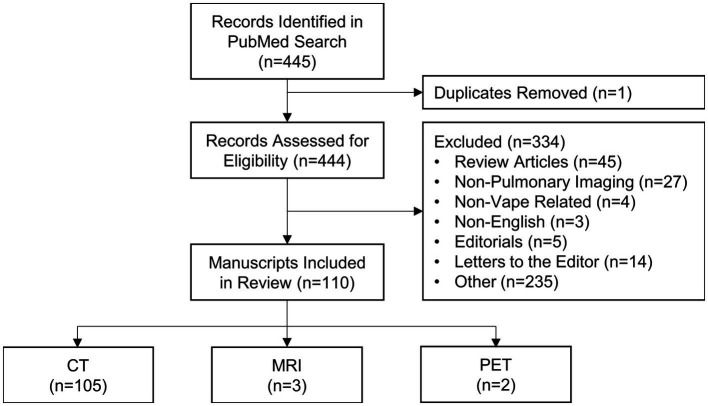
Methods: A systematic review was undertaken on June 13th, 2023 using PubMed to search for published manuscripts using the following criteria: [("Vaping" OR "e-cigarette" OR "EVALI") AND ("MRI" OR "CT" OR "Imaging")]. We included only studies involving human participants, vaping/e-cigarette use, and MRI, CT and/or PET.
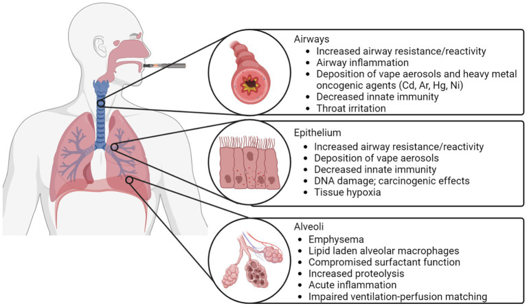
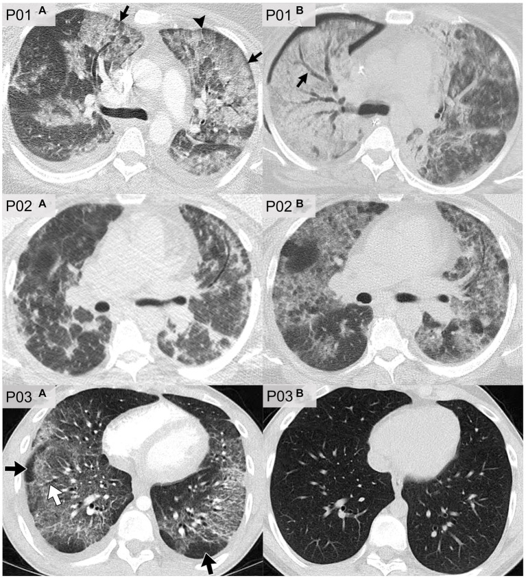
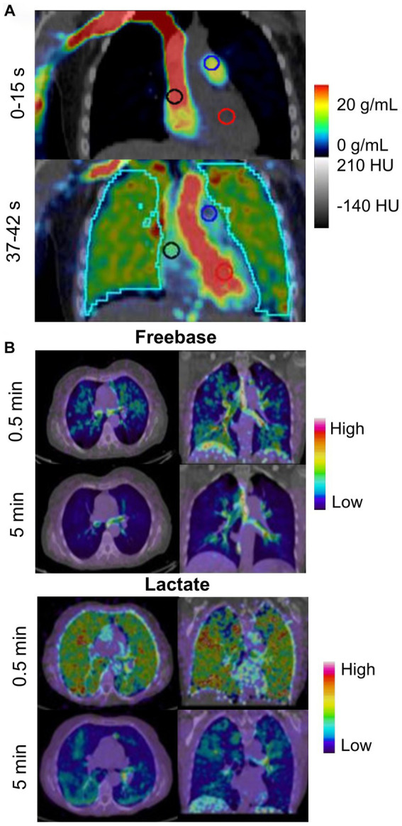
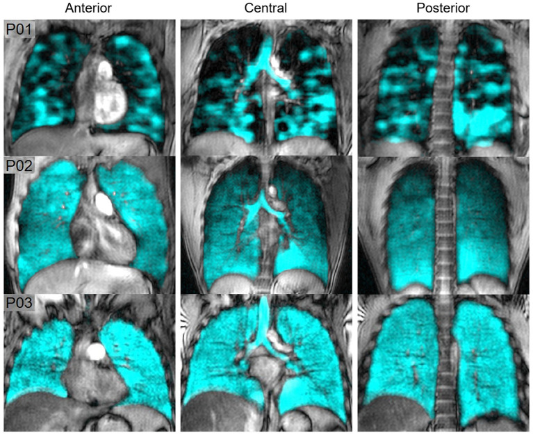
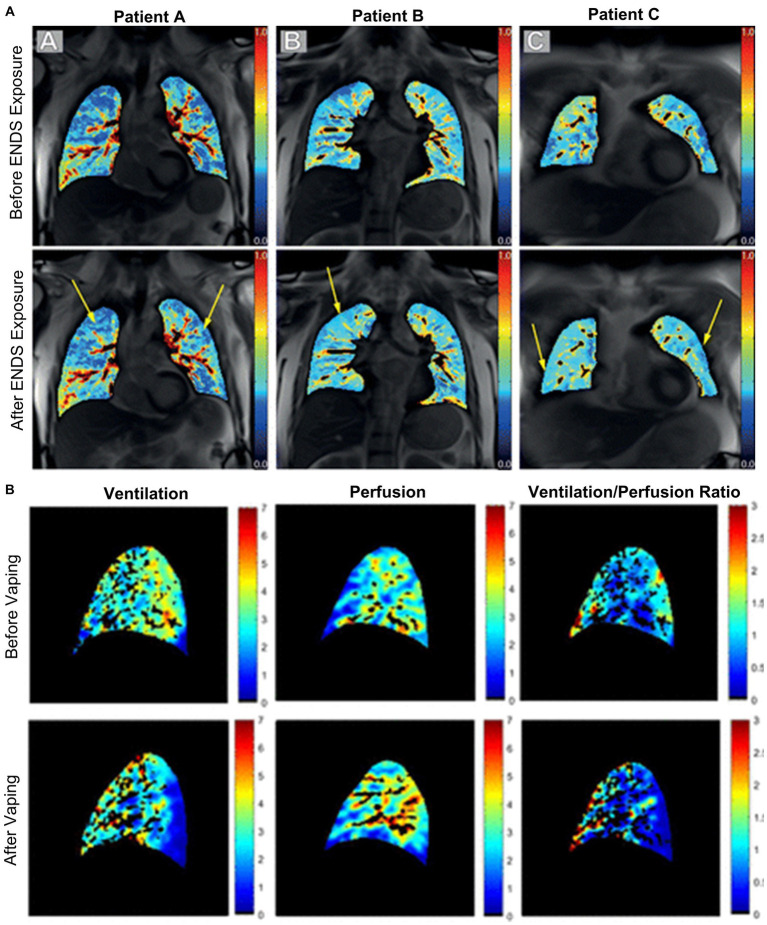
Results: The search identified 445 manuscripts, of which 110 (668 unique participants) specifically mentioned MRI, PET or CT imaging in cases or retrospective case series of patients who vaped. This included 105 manuscripts specific to CT (626 participants), three manuscripts which mainly used MRI (23 participants), and two manuscripts which described PET findings (20 participants). Most studies were conducted in North America (n = 90), with the remaining studies conducted in Europe (n = 15), Asia (n = 4) and South America (n = 1). The vast majority of publications described case studies (n = 93) and a few described larger retrospective or prospective studies (n = 17). In e-cigarette users and patients with EVALI, key CT findings included ground-glass opacities, consolidations and subpleural sparing, MRI revealed abnormal ventilation, perfusion and ventilation/perfusion matching, while PET showed evidence of pulmonary inflammation.
Discussion and conclusion: Pulmonary structural and functional imaging abnormalities were common in patients with EVALI and in e-cigarette users with or without respiratory symptoms, which suggests that functional MRI may be helpful in the investigation of the pulmonary health effects associated with e-cigarette use.
Keywords: CT; EVALI; MRI; PET; e-cigarettes.
Copyright © 2024 Hofmann, Poulos, Zhou, Sharma, Parraga and McIntosh.
Conflict of interest statement
GP discloses investigator initiated study funding from AstraZeneca, Novartis and GSK, study funding from the Ministry of Health and Long-term Care Ontario, and honoraria from AstraZeneca for speaking engagements, outside the submitted work. The remaining authors declare that the research was conducted in the absence of any commercial or financial relationships that could be construed as a potential conflict of interest. The reviewer LS declared a past co-authorship with the authors MS and GP to the handling editor.
Figures
Similar articles
-
E-cigarette, or vaping, product use-associated lung injury in adolescents: a review of imaging features.Pediatr Radiol. 2020 Mar;50(3):338-344. doi: 10.1007/s00247-019-04572-5. Epub 2020 Jan 2. Pediatr Radiol. 2020. PMID: 31897566
-
E-cigarette or vaping-associated lung injury (EVALI): a review of international case reports from outside the United States of America.Clin Toxicol (Phila). 2023 Feb;61(2):91-97. doi: 10.1080/15563650.2022.2160342. Epub 2023 Jan 13. Clin Toxicol (Phila). 2023. PMID: 36636876 Review.
-
CT Findings and Patterns of e-Cigarette or Vaping Product Use-Associated Lung Injury: A Multicenter Cohort of 160 Cases.Chest. 2021 Oct;160(4):1492-1511. doi: 10.1016/j.chest.2021.04.054. Epub 2021 May 3. Chest. 2021. PMID: 33957099 Free PMC article.
-
E-cigarette or vaping product use-associated lung injury in the pediatric population: imaging features at presentation and short-term follow-up.Pediatr Radiol. 2020 Aug;50(9):1231-1239. doi: 10.1007/s00247-020-04698-x. Epub 2020 Jun 3. Pediatr Radiol. 2020. PMID: 32495177
-
Radiological findings of e-cigarette or vaping product use associated lung injury: A systematic review.Heart Lung. 2021 Sep-Oct;50(5):736-741. doi: 10.1016/j.hrtlng.2021.05.004. Epub 2021 Jun 12. Heart Lung. 2021. PMID: 34130236
Cited by
-
E-Cigarette or Vaping Product Use-Associated Lung Injury: A Comprehensive Review.Int J Environ Res Public Health. 2025 May 17;22(5):792. doi: 10.3390/ijerph22050792. Int J Environ Res Public Health. 2025. PMID: 40427906 Free PMC article. Review.
-
A Systematic Literature Review on the Composition, Health Impacts, and Regulatory Dynamics of Vaping.Cureus. 2024 Aug 3;16(8):e66068. doi: 10.7759/cureus.66068. eCollection 2024 Aug. Cureus. 2024. PMID: 39229398 Free PMC article. Review.
References
-
- Siegel DA, Jatlaoui TC, Koumans EH, Kiernan EA, Layer M, Cates JE, et al. . Update: interim guidance for health care providers evaluating and caring for patients with suspected e-cigarette, or vaping, product use associated lung injury—United States, October 2019. MMWR Morb Mortal Wkly Rep. (2019) 68:919–27. doi: 10.15585/mmwr.mm6841e3, PMID: - DOI - PMC - PubMed
-
- Centers for Disease Control and Prevention . Outbreak of lung injury associated with the use of e-cigarette, or vaping, products. (2021). Available at: https://www.cdc.gov/tobacco/basic_information/e-cigarettes/severe-lung-d...
Publication types
LinkOut - more resources
Full Text Sources