[Preprint]. 2024 Jan 24:2024.01.23.576717.
doi: 10.1101/2024.01.23.576717.

Smad4 is essential for epiblast scaling and morphogenesis after implantation, but nonessential prior to implantation in the mouse

Affiliations

Smad4 is essential for epiblast scaling and morphogenesis after implantation, but nonessential prior to implantation in the mouse

Robin E Kruger et al. bioRxiv. .

Update in

Abstract

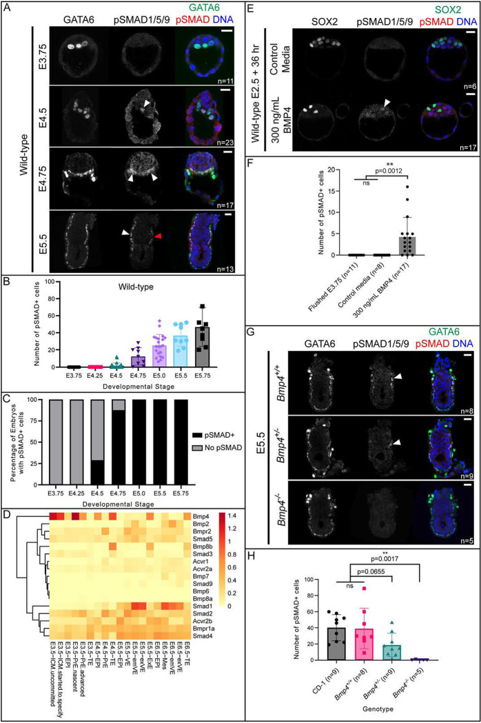
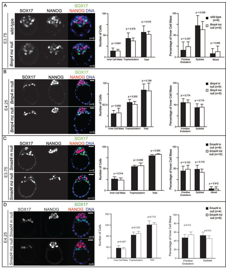
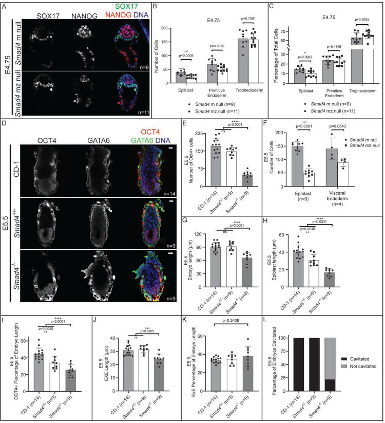
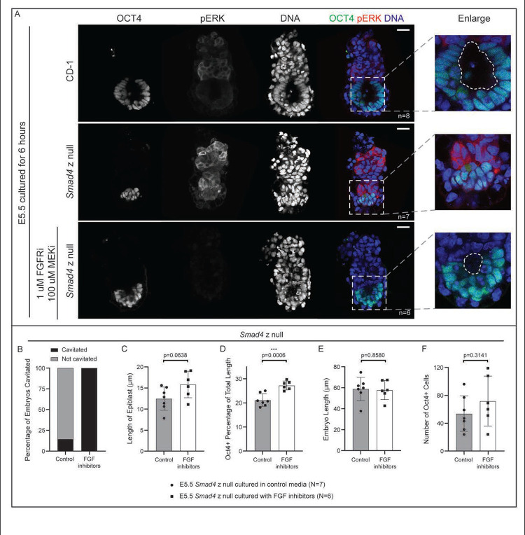
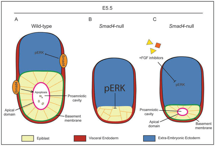
Bone Morphogenic Protein (BMP) signaling plays an essential and highly conserved role in axial patterning in embryos of many externally developing animal species. However, in mammalian embryos, which develop inside the mother, early development includes an additional stage known as preimplantation. During preimplantation, the epiblast lineage is segregated from the extraembryonic lineages that enable implantation and development in utero. Yet, the requirement for BMP signaling in mouse preimplantation is imprecisely defined. We show that, in contrast to prior reports, BMP signaling (as reported by SMAD1/5/9 phosphorylation) is not detectable until implantation, when it is detected in the primitive endoderm - an extraembryonic lineage. Moreover, preimplantation development appears normal following deletion of maternal and zygotic Smad4, an essential effector of BMP signaling. In fact, mice lacking maternal Smad4 are viable. Finally, we uncover a new requirement for zygotic Smad4 in epiblast scaling and cavitation immediately after implantation, via a mechanism involving FGFR/ERK attenuation. Altogether, our results demonstrate no role for BMP4/SMAD4 in the first lineage decisions during mouse development. Rather, multi-pathway signaling among embryonic and extraembryonic cell types drives epiblast morphogenesis post-implantation.

Keywords: epiblast; extraembryonic; maternal and zygotic gene deletion; morphogenesis; pSMAD1/5/9.


Conflict of interest statement

Competing interests The authors declare no competing interests.

Figures

Figure 1.
Figure 1.. BMP signaling becomes active in primitive endoderm at implantation.
A) SMAD1/5/9 phosphorylation (pSMAD1/5/9) in wild-type CD-1 embryos at E3.75, E4.5, E4.75, and E5.5. In all cases, positive pSMAD1/5/9 signal co-localizes with GATA6 as a marker of primitive endoderm and visceral endoderm. B) Quantification of total number of pSMAD1/5/9-positive cells in wild-type embryos in A and Supplemental Figure 1A. C) Quantification of the percentage of embryos from A and Supplemental Figure 1A which display any pSMAD1/5/9-positive cells versus no pSMAD1/5/9-positive cells. D) Heat map of the mean normalized expression of BMP pathway genes from scRNA-seq data from Nowotschin et al., 2019. E) pSMAD1/5/9 in wild-type embryos collected at E2.75 and cultured for 36 hours in media containing 300 ng/mL exogenous BMP4. F) Quantification of the total number of pSMAD1/5/9-positive cells in embryos from E revealed significantly more pSMAD1/5/9-positive cells in BMP4-treated embryos. G) pSMAD1/5/9 staining is absent in Bmp4 z null embryos at E5.5. H) Quantification of total number of pSMAD1/5/9-positive cells in wild-type and Bmp4-null embryos at E5.5 revealed significantly fewer pSMAD1/5/9-positive cells in Bmp4-null embryos. All pairwise comparisons were assessed by analysis of variance (ANOVA) with Tukey’s post-hoc test. White arrowheads indicate positive pSMAD1/5/9 signal. Red arrowhead indicates a GATA6+ cell which does not express pSMAD1/5/9. Scale bars represent 10 μm.
Figure 2.
Figure 2.. Maternal and zygotic Smad4 and Bmp4 are dispensable for blastocyst formation and preimplantation cell fate specification.
A) Immunofluorescence for SOX17 and NANOG as respective markers of primitive endoderm (PrE) and epiblast (EPI) in flushed E3.75 wild-type CD-1 embryos and embryos lacking maternal and zygotic Bmp4 (mz null). Quantification did not reveal any significant difference in cell number or cell fate between Bmp4 mz null embryos and controls. “Mixed” indicates co-expression of SOX17 and NANOG. B) Immunofluorescence for SOX17 and NANOG in flushed E4.25 embryos lacking maternal Bmp4 only (m null) and Bmp4 mz null embryos. Quantification did not reveal any significant difference in cell number or cell fate between Bmp4 mz null embryos and controls. “Mixed” indicates co-expression of SOX17 and NANOG. C) Immunofluorescence for SOX17 and NANOG in flushed E3.75 Smad4 m null and Smad4 mz null embryos. Quantification did not reveal any significant difference in cell number or cell fate between Smad4 mz null embryos and controls. D) Immunofluorescence for SOX17 and NANOG in flushed E4.25 Smad4 m null and Smad4 mz null embryos. Quantification did not reveal any significant difference in cell number or cell fate between Smad4 mz null embryos and controls. All pairwise comparisons were assessed by Student’s t-test. Scale bars represent 10 μm.
Figure 3.
Figure 3.. BMP-independent function of Smad4 is required for post-implantation epiblast organization and maintenance.
A) E4.75 Smad4 mz null embryos stained by immunofluorescence for SOX17 and NANOG. B) Quantification of EPI, PrE, and TE cell numbers from embryos in A revealed a significant decrease in EPI cells in Smad4 mz null embryos when compared to controls. C) Quantification of the EPI, PrE, and TE cells as a percentage of total cell number from embryos in A revealed a significant decrease in EPI percentage in Smad4 mz null embryos. D) E5.5 Smad4−/− embryos stained by immunofluorescence for OCT4 and GATA6 as markers of EPI and VE, respectively. Smad4−/− refers to combined Smad4 z null and Smad4 mz null embryos. E) Quantification of the number of OCT4+ cells in wild-type, Smad4+/−, and Smad4−/− embryos. F) Quantification of EPI and PrE cell numbers from Smad4+/− and Smad4−/− embryos at E5.5 revealed a specific, significant decrease in epiblast cell number in Smad4 mz null embryos when compared to controls (p<0.05 by Student’s t-test). The difference in VE cell numbers was not significant (p>0.05). G) Quantification of the proximal-distal length wild-type, Smad4+/−, and Smad4−/− embryos at E5.5. H) Quantification of the proximal-distal length of the EPI of wild-type, Smad4+/−, and Smad4−/− embryos at E5.5. I) Quantification of the proximal-distal length of the EPI as a percentage of total length of wild-type, Smad4+/−, and Smad4−/− embryos at E5.5. J) Quantification of the proximal-distal length of the EXE of wild-type, Smad4+/−, and Smad4−/− embryos at E5.5. K) Quantification of the proximal-distal length of the EXE as a percentage of total length of wild-type, Smad4+/−, and Smad4−/− embryos at E5.5. L) Quantification of the proportion of Smad4+/− and Smad4−/− embryos with a proamniotic cavity at E5.5. Comparisons in B, C, F were assessed by Student’s t-test. Comparisons in E, G-K were assessed by analysis of variance (ANOVA) with Tukey’s post-hoc test.
Figure 4.
Figure 4.. Inhibition of FGF signaling partially rescues epiblast cavitation in E5.5 Smad4 null embryos.
A) Wild-type and Smad4 z null embryos collected at E5.5 and cultured for 6 hours after dissection in control media or media containing FGFR/MEK inhibitors (see Methods), then stained by immunofluorescence for OCT4 and phosphorylated ERK (pERK). Dashed line in enlargement denotes the proamniotic cavity. B) Quantification of the proportion of treated and untreated Smad4−/− embryos with a proamniotic cavity at E5.5. C) Quantification of proximal-distal length of the EPI in treated and untreated E5.5 Smad4−/− embryos. D) Quantification of proximal-distal length of the EPI as a proportion of total length in treated and untreated E5.5 Smad4−/− embryos. E) Quantification of proximal-distal length in treated and untreated E5.5 Smad4−/− embryos. F) Quantification of OCT4-positive cell number in treated and untreated E5.5 Smad4−/− embryos.
Figure 5.
Figure 5.. FGF inhibition rescues rosette formation but not embryo growth in Smad4-null embryos.
A) In wild-type embryos, SMAD4 in the visceral endoderm promotes lumen formation in the epiblast by regulating a pro-apoptotic signal. Separately, SMAD4 also inhibits ERK phosphorylation in the EXE, which allows for polarization and rosette formation in the epiblast. B) In Smad4-null embryos, pERK is upregulated, causing an ectopic increase in pERK and preventing epiblast polarization. The pro-apoptotic signal is also lost. The combination of these two factors lead to epiblast disorganization and a failure to cavitate. C) Treatment with FGF inhibitors prevents upregulation of pERK in Smad4-null embryos. Repressed pERK levels allow epiblast polarization to proceed, resulting in a small proamniotic cavity.

References

    1. Bedzhov I. and Zernicka-Goetz M. (2014). Self-organizing properties of mouse pluripotent cells initiate morphogenesis upon implantation. Cell 156, 1032–1044. - PMC - PubMed
    1. Beppu H., Kawabata M., Hamamoto T., Chytil A., Minowa O., Noda T. and Miyazono K. (2000). BMP type II receptor is required for gastrulation and early development of mouse embryos. Developmental Biology 221, 249–258. - PubMed
    1. Blij S., Frum T., Akyol A., Fearon E. and Ralston A. (2012). Maternal Cdx2 is dispensable for mouse development. Development (Cambridge) 139, 3969–3972. - PMC - PubMed
    1. Boroviak T., Loos R., Lombard P., Okahara J., Behr R., Sasaki E., Nichols J., Smith A. and Bertone P. (2015). Lineage-Specific Profiling Delineates the Emergence and Progression of Naive Pluripotency in Mammalian Embryogenesis. Developmental Cell 35, 366–382. - PMC - PubMed
    1. Brennan J., Lu C.C., Norris D. P., Rodriguez T.A., Beddington R.S.P. and Robertson E.J. (2001). Nodal signalling in the epiblast patterns the early mouse embryo. Nature 411, 965–969. - PubMed

Publication types