

This is a preprint.
Cerebrovascular pathology mediates associations between hypoxemia during rapid eye movement sleep and medial temporal lobe structure and function in older adults
- PMID: 38328085
- PMCID: PMC10849660
- DOI: 10.1101/2024.01.28.577469
Cerebrovascular pathology mediates associations between hypoxemia during rapid eye movement sleep and medial temporal lobe structure and function in older adults
Abstract
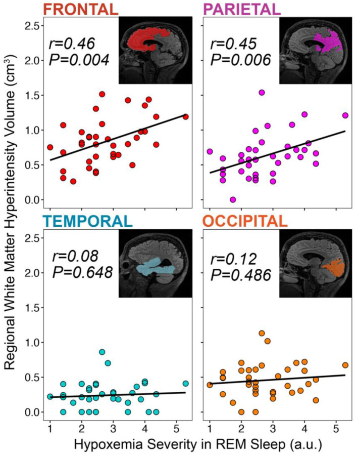
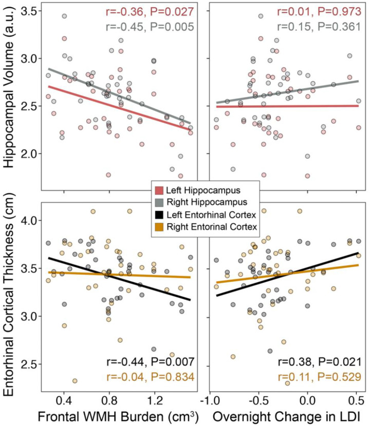
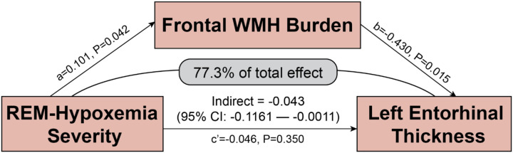
Obstructive sleep apnea (OSA) is common in older adults and is associated with medial temporal lobe (MTL) degeneration and memory decline in aging and Alzheimer's disease (AD). However, the underlying mechanisms linking OSA to MTL degeneration and impaired memory remains unclear. By combining magnetic resonance imaging (MRI) assessments of cerebrovascular pathology and MTL structure with clinical polysomnography and assessment of overnight emotional memory retention in older adults at risk for AD, cerebrovascular pathology in fronto-parietal brain regions was shown to statistically mediate the relationship between OSA-related hypoxemia, particularly during rapid eye movement (REM) sleep, and entorhinal cortical thickness. Reduced entorhinal cortical thickness was, in turn, associated with impaired overnight retention in mnemonic discrimination ability across emotional valences for high similarity lures. These findings identify cerebrovascular pathology as a contributing mechanism linking hypoxemia to MTL degeneration and impaired sleep-dependent memory in older adults.
Figures
Similar articles
-
Entorhinal Tau Pathology, Episodic Memory Decline, and Neurodegeneration in Aging.J Neurosci. 2018 Jan 17;38(3):530-543. doi: 10.1523/JNEUROSCI.2028-17.2017. Epub 2017 Nov 30. J Neurosci. 2018. PMID: 29192126 Free PMC article.
-
Reduced Repetition Suppression in Aging is Driven by Tau-Related Hyperactivity in Medial Temporal Lobe.J Neurosci. 2021 Apr 28;41(17):3917-3931. doi: 10.1523/JNEUROSCI.2504-20.2021. Epub 2021 Mar 17. J Neurosci. 2021. PMID: 33731446 Free PMC article.
-
Hypoxemia during rapid eye movement sleep mediates memory impairment in older adults at risk for dementia via CA1 hippocampal volume loss.Eur J Neurol. 2024 Dec;31(12):e16491. doi: 10.1111/ene.16491. Epub 2024 Sep 20. Eur J Neurol. 2024. PMID: 39302096 Free PMC article.
-
Evidence of neurodegeneration in obstructive sleep apnea: Relationship between obstructive sleep apnea and cognitive dysfunction in the elderly.J Neurosci Res. 2015 Dec;93(12):1778-94. doi: 10.1002/jnr.23634. Epub 2015 Aug 24. J Neurosci Res. 2015. PMID: 26301370 Review.
-
Obstructive sleep apnea, cognition and Alzheimer's disease: A systematic review integrating three decades of multidisciplinary research.Sleep Med Rev. 2020 Apr;50:101250. doi: 10.1016/j.smrv.2019.101250. Epub 2019 Dec 12. Sleep Med Rev. 2020. PMID: 31881487 Free PMC article.
References
-
- Slowik J. M., Sankari A. & Collen J. F. Obstructive Sleep Apnea. in StatPearls (StatPearls Publishing, Treasure Island (FL), 2023). - PubMed
Publication types
Grants and funding
LinkOut - more resources
Full Text Sources