[Preprint]. 2024 Jan 24:2024.01.23.576971.
doi: 10.1101/2024.01.23.576971.

Widespread Peptide Surfactants with Post-translational C-methylations Promote Bacterial Development

Affiliations

Widespread Peptide Surfactants with Post-translational C-methylations Promote Bacterial Development

Chen Zhang et al. bioRxiv. .

Update in

Abstract

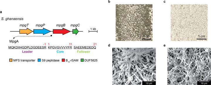
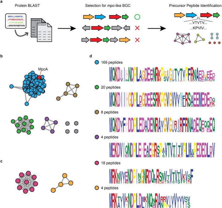
Bacteria produce a variety of peptides to mediate nutrient acquisition, microbial interactions, and other physiological processes. Of special interest are surface-active peptides that aid in growth and development. Herein, we report the structure and characterization of clavusporins, unusual and hydrophobic ribosomal peptides with multiple C-methylations at unactivated carbon centers, which help drastically reduce the surface tension of water and thereby aid in Streptomyces development. The peptides are synthesized by a previously uncharacterized protein superfamily, termed DUF5825, in conjunction with a vitamin B12-dependent radical S-adenosylmethionine metalloenzyme. The operon encoding clavusporin is wide-spread among actinomycete bacteria, suggesting a prevalent role for clavusporins as morphogens in erecting aerial hyphae and thereby advancing sporulation and proliferation.


Conflict of interest statement

Disclosures The authors declare no competing interests.

Figures

Extended Fig. 1
Extended Fig. 1
Structural elucidation of clavusporins. a-b, UPLC-coupled HR-MS analysis of extracts of WT S. clavuligerus and the deletion mutants indicated. Shown are extracted ion chromatograms of clavusporin A (a) and B (b). The traces are offset on the y-axis for clarity. c, Key NMR correlations used for determination of post-translational methylations in clavusporin B. d, Key NMR correlations used for determination of the overall structure of clavusporin B.
Extended Fig. 2
Extended Fig. 2
Phenotypic differences of WT S. clavuligerus and mpc mutants. a-c, Images of WT S. clavuligerus (a), ΔmpcB (b) and ΔmpcC (c) growing on GYM agar for 10 days. Note the green spores produced by the WT. d-f, SEM images of WT (d), ΔmpcB (e) and ΔmpcC (f), which confirm sporulation in WT, and minimal aerial hyphae growth in mutants. Each experiment was repeated in three biological replicates, and representative results are shown.
Extended Fig. 3
Extended Fig. 3
Phenotypic differences between WT S. ghanaensis and the mpgB mutant. a, The orthologous mpg gene cluster in S. ghanaensis. The gene locus of mpgA is marked by a black circle, and its protein sequence is shown. b-c, Images of WT S. ghanaensis (b) and ΔmpgB S. ghanaensis (c) growing on SFM agar for 3 days. Note the robust production of dark grey spores by the WT. d-e, SEM images of WT (d) and ΔmpgB S. ghanaensis (e), which show typical formation of Streptomyces spore chains in WT. Each experiment was repeated in three biological replicates, and representative results are shown.
Extended Fig. 4
Extended Fig. 4
MpcP is a site-specific leader protease. a, UPLC-HR-MS analysis of the MpcP reaction with NusA-MpcA. Heat-inactivated MpcP and a no-enzyme reaction were used as controls. Shown are extracted ion chromatograms of MpcA1–21. b, UPLC-HR-MS analysis of trypsin-digested NusA-MpcA as a positive control of MpcP assay. Shown are extracted ion chromatograms of two tryptic fragments of MpcA, MpcA1–13 (blue) and MpcA(−15)–(−1) (red). c, UPLC-HR-MS analysis of MpcP-treated MBP-MpcA fusion, which was co-expressed with MpcBC in E. coli. Shown are extracted ion chromatograms of MpcA1–21 with different methylation states. The traces are offset on the y-axis for clarity.
Fig. 1
Fig. 1
Discovery and structure of clavusporins. a, The mpc gene cluster as identified by genome mining. The gene locus of mpcA is marked by a black circle, and its protein sequence is shown. Strategies for genetic activation are shown below, including promoter exchange and codon optimization. b, MALDI-TOF MS analysis of SDS extracts of S. clavuligerus strains indicated. c, Numbering and parts of the MpcA peptide. d-e, MS/MS profiles of clavusporin A (d) and B (e). Blue and red arrows represent observed b and y ions, respectively. Each asterisk represents methylation on the corresponding residue. f, Structure of clavusporins. Post-translational methylations are marked in red.
Fig. 2
Fig. 2
Clavusporins reduce surface tension and promote aerial hyphae growth. a-b, Images of a water droplet consisting of 10% DMSO (a, vehicle control) and a droplet of clavusporin at 1 μg/μL in a 10% DMSO solution (b). c, Quantification of surface-tension reduction by clavusporins (red) and the unmethylated 13mer, MpcA1–13 (blue). Surface tension is linearly dependent on compound concentration. The lowest surface tension occurs at ~1 μg/μL clavusporins. d, Chemical complementation of S. clavuligerus ΔmpcBC with DMSO control, MpcA1–13, or clavusporins (10 μg dissolved in 2 μL of DMSO). e-g, SEM images of cell surfaces supplemented with DMSO control (e), MpcA1–13 (f), and clavusporins (g). Each experiment was repeated at least in three biological replicates, and representative results are shown.
Fig. 3
Fig. 3
Biosynthesis of clavusporins. a, Workflow for in vivo reconstitution of peptide methylations by co-expression of MBP-tagged precursor peptide and the modification enzymes. b-g, HPLC-coupled HR-MS analysis of MpcA core peptides from MBP-peptide fusions after column-enrichment and trypsin digestion. Shown are extracted ion chromatograms of MpcA1–13 (core peptide) with or without methylation, as indicated. The traces are offset on the y axis for clarity. h, Proposed biosynthetic pathway of clavusporins.
Fig. 4
Fig. 4
mpc-type BGCs are widespread in actinobacteria. a, Workflow for identification of mpc clusters and their putative precursor peptides. b-c, SSN of ‘long’ precursor peptides with acidic follower peptides (b) and ‘short’ precursor peptides without acidic follower peptides (c). Each node represents a unique BGC and the lines connecting them indicate sequence similarity in the precursor peptide (MpcA). The S. clavuligerus MpcA node is labeled in red. d, Logo plot for precursor peptides in each subfamily shown in b and c. The precursor peptides are color-coded to the BGCs in panels b and c, and the number of representatives for each cluster is shown.

Similar articles

References

    1. Clardy J. & Walsh C. Lessons from natural molecules. Nature 432, 829–837 (2004). - PubMed
    1. Salvador-Reyes L. A. & Luesch H. Biological targets and mechanisms of action of natural products from marine cyanobacteria. Nat. Prod. Rep. 32, 478–503 (2015). - PMC - PubMed
    1. Wang R. & Seyedsayamdost M. R. Hijacking exogenous signals to generate new secondary metabolites during symbiotic interactions. Nat. Rev. Chem. 1, 0021 (2017).
    1. van der Meij A., Worsley S. F., Hutchings M. I. & van Wezel G. P. Chemical ecology of antibiotic production by actinomycetes. FEMS Microbiol. Rev. 41, 392–416 (2017). - PubMed
    1. Bassler B. L. & Losick R. Bacterially speaking. Cell 125, 237–246 (2006). - PubMed

Publication types