[Preprint]. 2024 Jan 28:2024.01.27.577538.
doi: 10.1101/2024.01.27.577538.

Visualizing chaperone-mediated multistep assembly of the human 20S proteasome

Affiliations

Visualizing chaperone-mediated multistep assembly of the human 20S proteasome

Frank Adolf et al. bioRxiv. .

Update in

Abstract

Dedicated assembly factors orchestrate stepwise production of many molecular machines, including the 28-subunit proteasome core particle (CP) that mediates protein degradation. Here, we report cryo-EM reconstructions of seven recombinant human subcomplexes that visualize all five chaperones and the three active site propeptides across a wide swath of the assembly pathway. Comparison of these chaperone-bound intermediates and a matching mature CP reveals molecular mechanisms determining the order of successive subunit additions, and how proteasome subcomplexes and assembly factors structurally adapt upon progressive subunit incorporation to stabilize intermediates, facilitate the formation of subsequent intermediates, and ultimately rearrange to coordinate proteolytic activation with gated access to active sites. The structural findings reported here explain many previous biochemical and genetic observations. This work establishes a methodologic approach for structural analysis of multiprotein complex assembly intermediates, illuminates specific functions of assembly factors, and reveals conceptual principles underlying human proteasome biogenesis.

Keywords: 20S proteasome; PAC1; PAC2; PAC3; PAC4; POMP; chaperone; core particle; molecular machine; multiprotin complex; propeptide; protease; proteasome.


Conflict of interest statement

Competing Interest Statement J.W.H. is a founder and consultant for Caraway Therapeutics. B.A.S. is on the scientific advisory boards of Biotheryx and Proxygen. All other authors have no competing interests to declare.

Figures
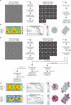
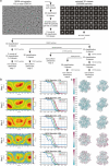
Ext. data figure 1.
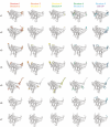
Ext. data figure 1.. Structure determination of recombinant human 20S proteasome
a, Cryo-EM image processing scheme for mature human 20S proteasome. b, Angular distribution and GSFSC curves of human 20S proteasome map. c, Local resolution of the cryo-EM map of human 20S proteasome map. d, Cryo-EM image processing scheme for preholo 20S CP and premature 20S CP. e, Angular distribution and GSFSC curves of preholo 20S CP (top) and premature 20S CP (bottom) maps. f, Local resolution of the cryo-EM maps of preholo 20S CP (top) and premature 20S CP (bottom) Scale bar in all micrographs represents 15 nm.
Ext. data figure 2.
Ext. data figure 2.. Structure determination of recombinant human proteasome assembly intermediates
a, Cryo-EM image processing scheme for the human proteasome assembly intermediates. b, Angular distribution and GSFSC curves of human proteasome assembly intermediates. c, Local resolution of the cryo-EM maps of human proteasome assembly intermediates. Scale bar in all micrographs represents 15 nm.
Ext. data figure 3.
Ext. data figure 3.. Structural comparison of human 20SCP assembly intermediates with yeast orthologs
a, Comparison of human 20S core particle with native substrate bound 26S proteasome (PDB: 6MSJ, only 20S core particle is shown). b, Comparison of human PAC3/4 (yellow) with yeast Pba3/4 (green, PDB:2Z5C) reveals their structural similarity. c, Comparison of structure 1 and structure 5 shows the position of PAC3/4 in structure 1 is occupied by β4–7 later in structure 5. d, Comparison of human β2 (pink) with yeast β2 (green, PDB:7LS6/1RYP) in pre-15S (top) and mature 20S CP (bottom) shows the insertion of yeast α2 changes the trajectory of yeast β2 C-terminal loop compared to human in immature 20S CP. e, Comparison of human PAC1/2 (red) with yeast Pba1/2 (green, PDB:7LSX) shows the N-terminus of PAC1 and Pba1 interacts with human α1 and yeast α2, respectively, and make close contact with POMP/Ump1. f, Comparison of structure 1 POMP interaction with PAC3 and structure 5 POMP interaction with β5 propeptide
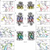
Ext. data figure 4.
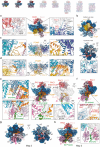
Ext. data figure 4.. Fusion-tetrad formed by the opposing β subunits upon half CP fusion
a, Cryo-EM map of structure 5 with β-subunits colored in yellow. b, Cryo-EM map of preholo CP with β-subunits of one half CP colored in purple and the other half CP colored in salmon. c-f, Close-up view of fusion-tetrad between β3 (structure 5 in yellow and preholo 20SCP in purple) and β5 from the opposite half CP (salmon) (c), two opposing β4 (d), β6 and β2 from the opposite half CP (e) and β7 and β1 from the opposite half CP (f). g, Cryo-EM map of mature 20S CP with β-subunits of one half CP colored in green and the other half CP colored in blue. h-k, Close-up view of fusion-tetrad between β2 (preholo 20SCP in purple and mature 20SCP in green) and β5 from the opposite half CP (preholo 20SCP in salmon and mature 20SCP in blue) (h-i), β5 and β3 from the opposite half CP (j), and β1 and β7 from the opposite half CP (k).
Ext. data figure 5.
Ext. data figure 5.
Structures of assembly chaperons and β subunits across β-ring formation
Ext. data figure 6.
Ext. data figure 6.
Structural comparison of α subunits across β-ring formation
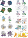
Fig. 1.
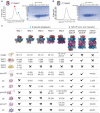
Fig. 1.. Recombinant expression of human 20S proteasome in insect cells
a, Purification of recombinant β2-tagged (PSMB7) 20S CP. Eluates were subjected to size exclusion chromatography (SEC) (right panel), and the fractions analyzed by SDS polyacrylamide gel electrophoresis (SDS-PAGE) followed by Coomassie staining (left panel). Asterisk indicates novel bands in the later fractions that likely correspond to the assembly chaperones, PAC1–4 and POMP. Red dot indicates main peak fractions corresponding to half CP fused 20SCP and blue dot indicates shoulder peak fractions corresponding to 20SCPassembly intermediates. b, Purification of recombinant β7-tagged (PSMB4) 20S CP. Eluates were subjected to size exclusion chromatography (right panel), and the fractions analyzed by SDS polyacrylamide gel electrophoresis (SDS-PAGE) followed by Coomassie staining (left panel). c, Cryo-EM maps of eight distinct CP subcomplexes. Their subunits, chaperone configurations, and the resolved region in each map are indicated.
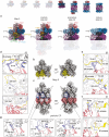
Fig. 2.
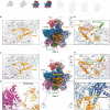
Fig. 2.. Structural progression of early assembly intermediates
a, Cross-section view of cryo-EM map of the structure 1 shows all five assembly chaperons. PAC1/2 is perched on top of the α-ring and interact with all α-subunits except for α3. b, Bottom-up view (top panel) of cryo-EM map of structure 1 shows that PAC3/4 resides in the groove between α4/5 and α5/6, and β2 sits in the groove between α1 and α2. Top-down (bottom panel) of structure 1 shows that PAC1/2 holds the α-ring ‘gate’ open and the pore is open. c, Side view of structure 1 shows that PAC3/4 interacts with α3-α7 of α-ring. d, Side view of structure 1 shows that POMP interacts with α1- α3 and α7 of α-ring. e, Cross-section view of cryo-EM map of structure 2 shows incorporation of the β3 is mutually stabilized by β2-propeptide and β2 C-terminal extension (CTE). Close-up view shows additional N-terminal region of POMP interacting with β2-propeptide and β3. f, Close-up view of PAC1 N-terminus of structure 2 shows its interaction with α3 and POMP. g, Bottom-up view (left panel) of cryo-EM map of structure 2 shows that release of PAC3/4 frees up α4/5 and α5/6 grooves and β3 occupies α2/3 groove. Top-down view (right panel) shows that the pore is closed. h, Cross-section view of cryo-EM map of structure 3 shows incorporation of the β4 and its interaction with β3 and the α-ring.
Fig. 3.
Fig. 3.. Distinct structural features upon β5 and β6 incorporation
a-b, Cross-section view of cryo-EM map of the structure 3 shows PAC1/2, POMP and β2-β4. Close-up view of POMP (a) shows its helix-turn-helix configuration and its interaction with the α-ring and β-subunits. Close-up view (b) reveals interaction between POMP and β2-propeptide. c-f, Cross-section view of cryo-EM map of the structure 4 shows PAC1/2, POMP and β2-β5 (β6 is cropped in the cross-section) upon β5 and β6 incorporation. Close-up view of POMP (c) shows that additional N-terminal region of POMP (blue) is resolved and stabilizes β5 and β6 incorporation. Close-up view of β2-propeptide (d) reveals the additional resolved N-terminal region of POMP interacts with the N-terminus of β2-propeptide and drives it to a different orientation. Close-up view of β5, β6 and POMP (e) shows that the additional resolved N-terminal region of POMP interacts with β5 loop. Close-up view of β5-propeptides (f) shows its interaction with α6, α7, α1 and POMP.
Fig. 4.
Fig. 4.. Incorporation of β7 and β1 completes β-ring and reveals all β-propeptides
a-b, Cross-section view of cryo-EM map of the structure 4 shows PAC1/2, POMP, β3 and β6 (β2 is hidden behind and β4–5 are cropped in the cross-section) upon β5 and β6 incorporation. Close-up view of β-ring (a) and interaction between β5-propeptide and POMP (b). c-f, Cross-section view of cryo-EM map of the structure 5 shows PAC1/2, POMP, β3 and β6 (β7 and β1–2 are hidden behind and β4–5 are cropped in the cross-section) upon β7 and β1 incorporation. Close-up (c) shows additional β5-propeptide is resolved upon completion of the β-ring (blue) compare to β5 in Structure 4. Close-up view of β5-propeptide’s interaction with POMP, β1 and β2 (d) shows that POMP’s N-terminus (blue) is resolved upon completion of the β-ring and interacts with β5-propeptide. Close-up view of newly incorporated β1 and β7 (e) reveals the β1-propeptide and its interaction with β7 N-terminus and loop. Close-up view (f) of β5-propeptide’s interaction with α5–7, β5–6 and β1.
Fig. 5.
Fig. 5.. Half CP fusion and 20SCP maturation
a, Cross-section view of cryo-EM map of structure 5, preholo-20SCP, premature-20SCP and mature 20SCP shows the presence and release of assembly chaperones and β-propeptides during 20SCP fusion and maturation process. b, Cryo-EM map of structure 5 with β1, β2 and β5 colored in yellow. c, Cryo-EM map of preholo CP with β1, β2 and β5 of one half CP colored in purple and the other half CP colored in salmon. d, Close-up view of β2 structural changes (β2 from structure 5 colored in yellow and β2 from preholo CP colored in purple) and its interaction with β6 (salmon) from the opposite half CP upon CP fusion. e, Close-up view of β5 structural changes (β5 from structure 5 colored in yellow and β5 from preholo CP colored in purple) and its interaction with β3 (salmon) from the opposite half CP upon CP fusion. f, Close-up view of β1 structural changes (β1 from structure 5 colored in yellow and β1 from preholo CP colored in purple) and its interaction with β7 (salmon) from the opposite half CP upon CP fusion. g, Close-up view of fusion-tetrad formed by the fusion-hairpin and fusion-loop from the opposing β subunits.
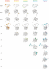
Fig. 6.
Fig. 6.. Schematic representation of chaperon-mediated stepwise assembly of the 20S CP and summary of high-resolution structures of human assembly intermediates.
Assembly of the 20S core particle can be distinguished into the three steps: (i) α-ring assembly, (ii) β-ring assembly, and (iii) half CP fusion and maturation. High resolution maps of human CP assembly intermediated presented in this study are indicated below their schematic representations

References

    1. Lowe J., Stock D., Jap B., Zwickl P., Baumeister W. & Huber R. Crystal structure of the 20S proteasome from the archaeon T. acidophilum at 3.4 A resolution. Science 268, 533–539 (1995). 10.1126/science.7725097 - DOI - PubMed
    1. Groll M. et al. Structure of 20S proteasome from yeast at 2.4 A resolution. Nature 386, 463–471 (1997). 10.1038/386463a0 - DOI - PubMed
    1. Groll M. et al. A gated channel into the proteasome core particle. Nat Struct Biol 7, 1062–1067 (2000). 10.1038/80992 - DOI - PubMed
    1. Unno M. et al. The structure of the mammalian 20S proteasome at 2.75 A resolution. Structure 10, 609–618 (2002). 10.1016/s0969-2126(02)00748-7 - DOI - PubMed
    1. Arendt C. S. & Hochstrasser M. Identification of the yeast 20S proteasome catalytic centers and subunit interactions required for active-site formation. Proc Natl Acad Sci U S A 94, 7156–7161 (1997). 10.1073/pnas.94.14.7156 - DOI - PMC - PubMed

Publication types