

Supratrochlear Rim is Correlated with Isolated Patellar Chondromalacia on Magnetic Resonance Imaging of the Knee
- PMID: 38328532
- PMCID: PMC10847026
- DOI: 10.1016/j.asmr.2023.100855
Supratrochlear Rim is Correlated with Isolated Patellar Chondromalacia on Magnetic Resonance Imaging of the Knee
Abstract
Purpose: To investigate the relationship between the supratrochlear rim and isolated patellar chondromalacia (PC) using magnetic resonance imaging (MRI) scans of the knee.
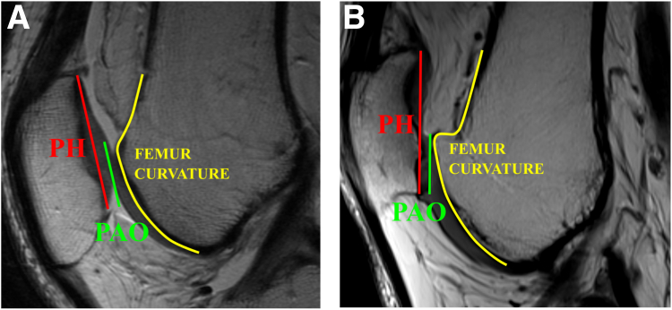
Methods: Patients without patellofemoral pain (control group) and patients with patellofemoral pain and diagnosed with stage III or IV PC based on MRI (defect group) were retrospectively identified. Patients with a history of patellar subluxation were excluded. We used patient MRI scans to perform 20 anatomical measurements of the patellofemoral joint. We also performed 2 measurements of the anterior femoral curvature. A total of 30 patients (29 ± 8.7 years) were in the control group, and 20 patients were in the defect group (29.4 ± 9.7 years).
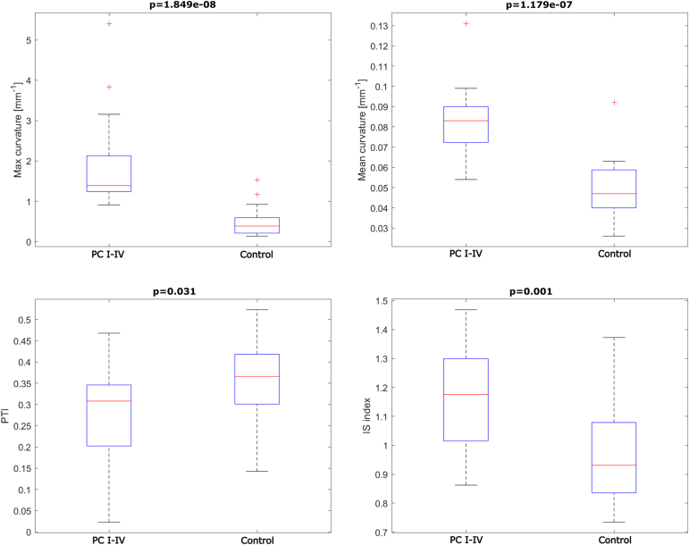
Results: The maximum curvature (P < .001) and mean curvature (P < .001) of the anterior femoral condyle were found statistically significantly different between the groups. Patellotrochlear index (P = .03) and Insall-Salvati index (P < .001) were also found statistically significantly different between the 2 groups. Patella type III and trochlear dysplasia grade B were found more common in the defect group.
Conclusions: In this Level III prognostic, case-control study, we have shown through MRI knee measurements that the isolated patellar chondromalacia in patients without a history of patellar subluxation and dislocation is correlated with the increased anterior femoral curvature in combination with patella alta.
© 2023 The Authors.
Conflict of interest statement
The authors declare the following financial interests/personal relationships, which may be considered as potential competing interests: T.P. reports personal fees from Rehasport Clinic outside the submitted work. Full ICMJE author disclosure forms are available for this article online, as supplementary material.
Figures


References
-
- Heintjes E.M., Berger M., Bierma-Zeinstra S.M., et al. Exercise therapy for patellofemoral pain syndrome. Cochrane Database Syst Rev. 2003;4:CD003472. - PubMed
-
- Mølgaard C., Rathleff M.S., Simonsen O. Patellofemoral pain syndrome and its association with hip, ankle, and foot function in 16- to 18-year-old high school students: a single-blind case-control study. J Am Podiatr Med Assoc. 2011;101:215–222. - PubMed
-
- Fairbank J., Pynsent P., van Poortvliet J.A., Phillips H. Mechanical factors in the incidence of knee pain in adolescents and young adults. J Bone Joint Surg Br. 1984;66:685–693. - PubMed
-
- Wood L., Muller S., Peat G. The epidemiology of patellofemoral disorders in adulthood: A review of routine general practice morbidity recording. Prim Health Care Res Dev. 2011;12:157–164. - PubMed
Grants and funding
LinkOut - more resources
Full Text Sources
