. 2024 Feb 7;3(2):pgae018.
doi: 10.1093/pnasnexus/pgae018. eCollection 2024 Feb.

p17/C18-ceramide-mediated mitophagy is an endogenous neuroprotective response in preclinical and clinical brain injury

Affiliations

p17/C18-ceramide-mediated mitophagy is an endogenous neuroprotective response in preclinical and clinical brain injury

Eda Karakaya et al. PNAS Nexus. .

Abstract

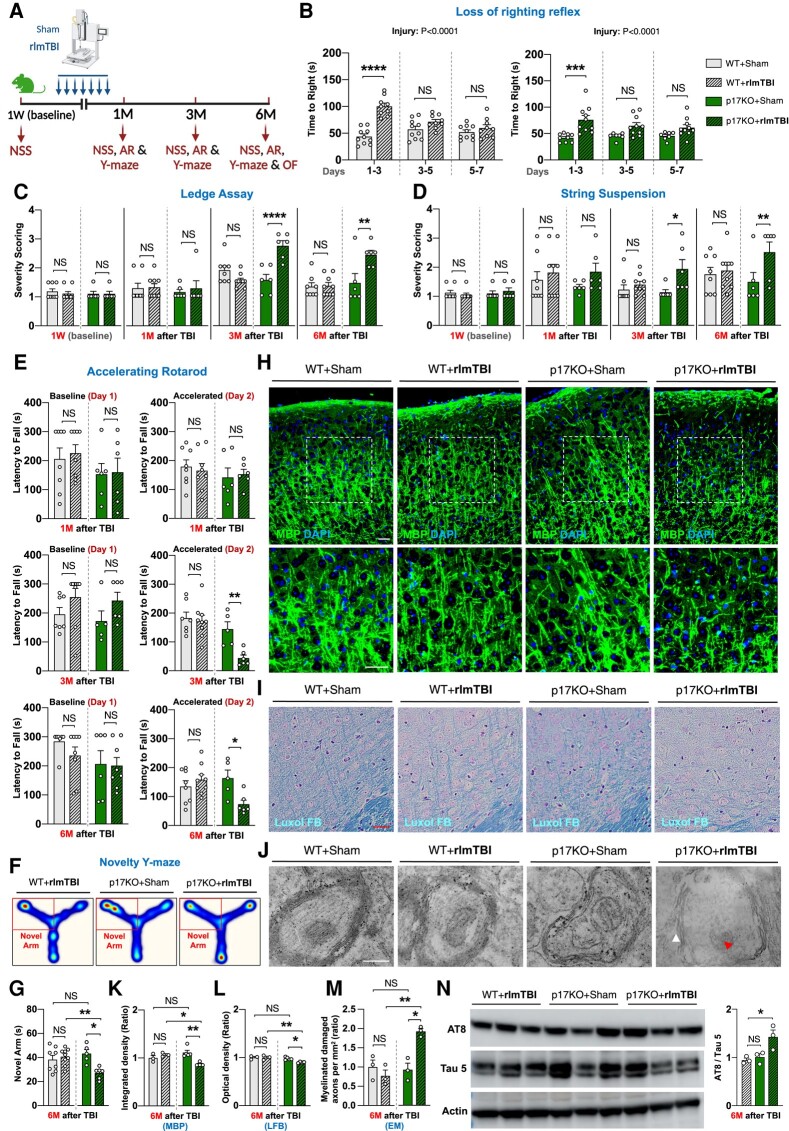
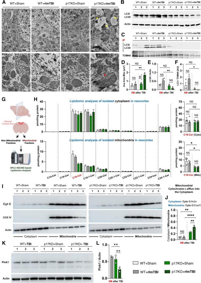
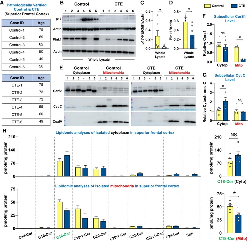
Repeat concussions (or repetitive mild traumatic brain injury [rmTBI]) are complex pathological processes consisting of a primary insult and long-term secondary complications and are also a prerequisite for chronic traumatic encephalopathy (CTE). Recent evidence implies a significant role of autophagy-mediated dysfunctional mitochondrial clearance, mitophagy, in the cascade of secondary deleterious events resulting from TBI. C18-ceramide, a bioactive sphingolipid produced in response to cell stress and damage, and its synthesizing enzyme (CerS1) are precursors to selective stress-mediated mitophagy. A transporter, p17, mediates the trafficking of CerS1, induces C18-ceramide synthesis in the mitochondrial membrane, and acts as an elimination signal in cell survival. Whether p17-mediated mitophagy occurs in the brain and plays a causal role in mitochondrial quality control in secondary disease development after rmTBI are unknown. Using a novel repetitive less-than-mild TBI (rlmTBI) injury paradigm, ablation of mitochondrial p17/C18-ceramide trafficking in p17 knockout (KO) mice results in a loss of C18-ceramide-induced mitophagy, which contributes to susceptibility and recovery from long-term secondary complications associated with rlmTBI. Using a ceramide analog with lipid-selenium conjugate drug, LCL768 restored mitophagy and reduced long-term secondary complications, improving cognitive deficits in rlmTBI-induced p17KO mice. We obtained a significant reduction of p17 expression and a considerable decrease of CerS1 and C18-ceramide levels in cortical mitochondria of CTE human brains compared with age-matched control brains. These data demonstrated that p17/C18-ceramide trafficking is an endogenous neuroprotective mitochondrial stress response following rlmTBI, thus suggesting a novel prospective strategy to interrupt the CTE consequences of concussive TBI.


Figures

Fig. 1.
Fig. 1.
The mouse model of new repetitive mild closed head injury demonstrates age-at-injury–dependent secondary neuropathological changes and cognitive impairments. A) Experimental setup. Two-month-old (2M) and 12-month-old (12M) male C57BL/6J WT mice underwent rlmTBIs or sham injuries (Created with Biorender.com), and (B) the latency of their righting reflex was recorded, followed by functional and pathological examination for 8 months. The long-term effects of the novel rlmTBI on neurological deficits in both the 2M and 12M mice groups were longitudinally assessed by (C) the Ledge assay (LA) in mice before rlmTBI and 1 and 8 months after the last injury. Cognitive performance was assessed by Barnes maze (BM) in 2M (D) and 12M (E) mice; escape latency across the 5 days of acquisition phase (left) and two consecutive sessions (right, 5 min interval) on day 3 of the acquisition phase; and movement heat map on days 1 and 3 (the occupancy rate is graded by a color map ranging from cold to warm colors). F and G) Anxiety-like behavior was assessed by bright light, open-field (OF) in both groups; time spent in the center (left) and distance traveled (right) and trajectory plots at 1 and 8 months after the last injury. The long-term neuropathological consequences of rlmTBI of the 2M and 12M mice groups on the axonal degeneration (axonopathy) and tau pathology, as shown by immunofluorescence (IF) for MBP (H) and LFB staining (I) to myelinated axonopathy and Bielschowsky silver staining (J) to disruption of axons and the presence of axonal swellings and spheroids (red arrow heads indicate axonal swellings; white arrow heads indicate spheroids) and immunoblotting for AT180, AT8, and Tau5 (total tau) (K) to abnormal tau phosphorylation in neocortex and corpus callosum. Inset images are the high magnification images of the selected area denoted by white. Scale bar, 50 μm. Data are expressed as mean ± SEM (two-way ANOVA with Bonferroni's correction and unpaired two-tailed nonparametric t test). NS, not significant. *P < 0.05, **P < 0.01, ***P < 0.001, and ****P < 0.0001.
Fig. 2.
Fig. 2.
Ablation of p17 affects the long-term functional recovery and development of secondary axonal degeneration after rlmTBI. A) Experimental setup. Two-month-old male p17KO and C57BL/6J WT mice underwent rlmTBIs or sham injuries, and (B) the latency of their righting reflex was recorded, followed by functional and pathological examination for 6 months. Their neurological scoring (NSS) was longitudinally assessed by (C) Ledge assay and (D) string suspension test in mice before TBI and 1, 3, and 6 months after the last injury. E) Sensorimotor competency was longitudinally assessed with the accelerated rotarod (AR) in mice 1, 3, and 6 months after the last injury (day 1 represents baseline performance; maximum time is 300 s). Working memory and cognitive flexibility were assessed by novelty Y-maze (F), movement heat map (the occupancy rate is graded by a color map ranging from cold to warm colors), and (G) cumulative time spent in novel arm on test phase. The microstructural organization of myelinated axons and axonal integrity were assessed by immunostaining for (H) MBP, (I) LFB myelin staining, and (J) electron microscopy (red arrow head indicate degenerated axonal mitochondria; white arrow head indicate disorganized myelin attachment to axons at paranodes; scale bar, 500 nm) in the neocortex 6 months after the last injury. Inset images are high magnifications of representative areas. Bar graph showing the quantification of integrated fluorescence density of (K) MBP immunostaining and optical density of (L) LFB staining in the neocortex of mice in each group (n = 3–5). Scale bars, 50 μm. M) Bar graph showing the quantification of the relative myelinated damaged axons at electron microscopy (EM) imagining in the neocortex of mice in each group (n = 3). N) Western blot analysis of cortical tau pathology was analyzed for AT8, AT100, and Tau5 (total tau) at 6 months after rlmTBIs (n = 3). Data are expressed as mean ± SEM (two-way ANOVA with Bonferroni's correction and one-way ANOVA with Dunnett's correction). NS, not significant. *P < 0.05, **P < 0.01, ***P < 0.001, and ****P < 0.0001.
Fig. 3.
Fig. 3.
The accumulation of dysfunctional mitochondria due to impaired p17/C18-Cer–associated mitophagy contributes to secondary axonal degeneration after rlmTBI. A new cohort of 2-month-old male p17KO and C57BL/6J WT mice underwent rlmTBIs or sham injuries, followed by the analysis of mitochondrial function, structure, and intracellular organization at 6-month post-rlmTBIs. A) Ultrastructural pathologies of axonal mitochondria of the cortical neurons and (D) quantification (average area of the mitochondria) were determined. The red arrow head indicates tubular and large mitochondria that are likely to represent fusion at early stages of myelinated axonal mitochondria; the yellow arrow heads indicate autophagic vacuoles and engulfed mitochondria; the white arrow head indicates darkened nucleoplasm and a crenated nucleolemma of the oligodendrocyte. The below images exhibited the higher magnification of each corresponding dashed boxed area in the upper panels. Scale bars, 1 μm. B and E) The lipidated LC3 (LC3-II) was analyzed with immunoblotting in the neocortex. C and F) Using co-IP analysis, the association between LC3/TOM40 was analyzed to measure mitophagy. G) A schematic representation of the microdissection procedure used to isolate the affected cortical region. H) HPLC-MS/MS–based lipidomics analyses of bioactive ceramide profiles in mitochondrial and nonmitochondrial fractions isolated from pathologically relevant cortical regions. I and J) Mitochondrial and cytosolic Cytochrome c protein levels were analyzed with immunoblotting in the neocortex with anti-Cytochrome c antibody. Whole-cell lysates were analyzed for (K and L) PINK1 expression by immunoblotting in the neocortex. Loading standards were actin for homogenate and Cox IV for mitochondria (n = 3–5). Data are expressed as mean ± SEM (two-way ANOVA with Bonferroni's correction and one-way ANOVA with Dunnett's correction). NS, not significant. *P < 0.05, **P < 0.01, and ****P < 0.0001.
Fig. 4.
Fig. 4.
C18-Cer analog, LCL768, restores mitophagy and reduces secondary disease development in p17KO mice after rlmTBI. A) The chemical structure of LCL768 contains two C18-Pyr-Cer moieties conjugated with selenium. B) Experimental setup. Two-month-old male p17KO mice underwent rlmTBIs or sham injuries, and 1 day after the last injury, they were chronically treated with LCL768 (0.1 mg/kg) or vehicle (PBS) through intracranially (inserted directly into the overlaying cortex) implanted osmotic minipumps (Alzet) for 7 weeks. Treatment began 1 day after their last injury and continued for 7 consecutive weeks with 3 months of washout. C) The loss of the righting reflex was evaluated based on the latency of self-righting immediately after the injuries. Their neurological competency (NSS and AR) was longitudinally assessed by (D) Ledge assay, (E) string suspension, and (F) accelerated rotarod tests in mice before injury and 6 months after the last injury. (G) Working memory performance was assessed with the novelty Y-maze in mice 6 months after the last injury. The microstructural organization of myelinated axons and axonal integrity were assessed by immunostaining for (H) LFB, (J) MBP myelin, and (I) Bielschowsky silver staining. Bar graph showing the quantification of optical density of LFB staining and integrated fluorescence density of MBP immunostaining in the neocortex of mice in each group (n = 3). Scale bars, 50 μm. K and L) Western blot analysis of cortical tau pathology was analyzed for AT8, AT180, and Tau5 (total tau) at 6 months after rlmTBIs (n = 4–5). M) AT8-positive pathological tau was also assessed by immunohistology (n = 3). N) Electron microscopy (red arrows indicate degenerated axonal mitochondria; white arrows indicate disorganized myelin attachment to axons at paranodes; Scale bar, 500 nm) in the neocortex 6 months after the last injury. Inset images are high magnifications of representative areas. Bar graph showing the quantification of the relative (O) myelinated damaged axons and (P) axons with swollen mitochondria at EM imagining in the neocortex of mice in each group (n = 3). Data are expressed as mean ± SEM (one-way ANOVA with Dunnett's correction). NS, not significant. *P < 0.05 and **P < 0.01.
Fig. 5.
Fig. 5.
Significant alterations of the p17/CerS1–mitophagy axis in patients with CTE. A) The six neuropathologically verified male CTE brain specimens of the superior frontal cortex under the age of 75 and six age-matched healthy controls were subjected to immunoblot and lipidomic analysis of subcellular fraction. Whole-cell lysates were analyzed for (B and C) p17 and (B and D) PINK1 expressions by immunoblotting in the superior frontal cortex. Mitochondrial and cytosolic (E and F) CerS1 and (E and G) Cytochrome c protein levels were analyzed with immunoblotting in the neocortex with anti-Cytochrome c and CerS1 antibodies, respectively. H) Mass spectrophotometry–based lipidomics analyses of cytoplasmic and mitochondrial C14–C26-ceramide levels in the superior frontal cortex. Loading standards were actin for homogenate and Cox IV for mitochondria (n = 6). Data are expressed as mean ± SEM (two-way ANOVA with Bonferroni's correction). NS, not significant. *P < 0.05 and **P < 0.01.
Fig. 6.
Fig. 6.
p17/C18-Cer–mediated mitophagy is an endogenous neuroprotective mitochondrial stress response at the time of brain injury and beyond. Subcellular localization of CerS1 by the novel p17 transporter in damaged mitochondria vs. ER to induce C18-Cer generation, and the subsequent injury/stress requires LC3 activation and mitophagy in various metabolically active tissues, including the brain.

References

    1. McKee AC, et al. 2013. The spectrum of disease in chronic traumatic encephalopathy. Brain. 136(Pt 1):43–64. - PMC - PubMed
    1. Smith DH, Johnson VE, Stewart W. 2013. Chronic neuropathologies of single and repetitive TBI: substrates of dementia? Nat Rev Neurol. 9(4):211–221. - PMC - PubMed
    1. Lye TC, Shores EA. 2000. Traumatic brain injury as a risk factor for Alzheimer's disease: a review. Neuropsychol Rev. 10(2):115–129. - PubMed
    1. Nordstrom P, Michaelsson K, Gustafson Y, Nordstrom A. 2014. Traumatic brain injury and young onset dementia: a nationwide cohort study. Ann Neurol. 75(3):374–381. - PubMed
    1. Coronado VG, et al. 2011. Surveillance for traumatic brain injury-related deaths–United States, 1997–2007. MMWR Surveill Summ. 60(5):1–32. - PubMed