. 2024 Feb 8;9(3):e168443.
doi: 10.1172/jci.insight.168443.

Abnormalities in microbiota/butyrate/FFAR3 signaling in aging gut impair brain function

Affiliations

Abnormalities in microbiota/butyrate/FFAR3 signaling in aging gut impair brain function

Sidharth P Mishra et al. JCI Insight. .

Abstract

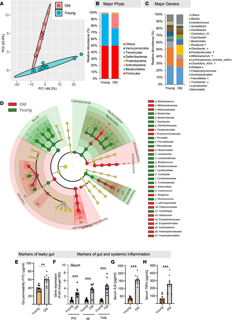
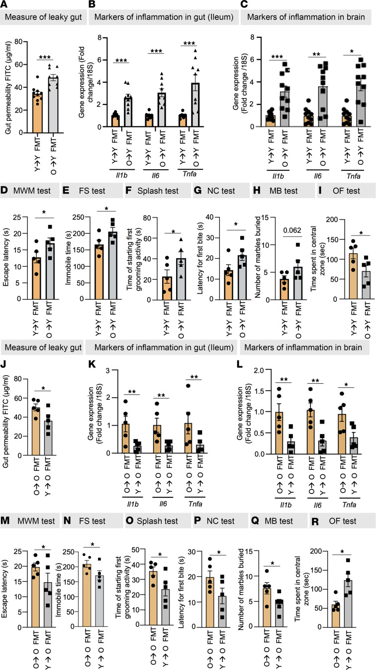
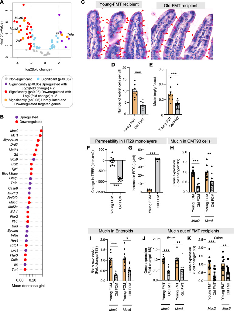
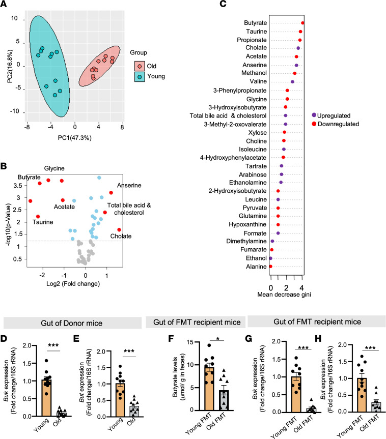
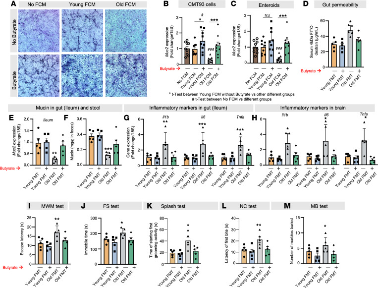
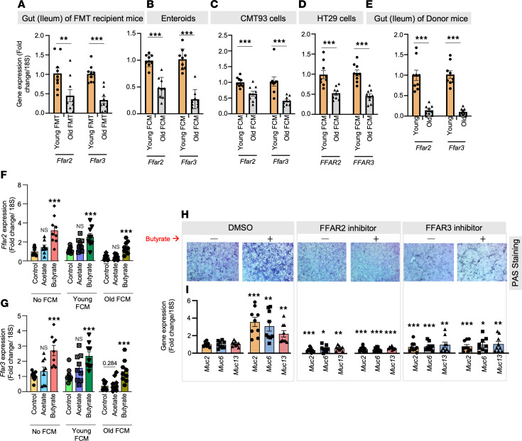
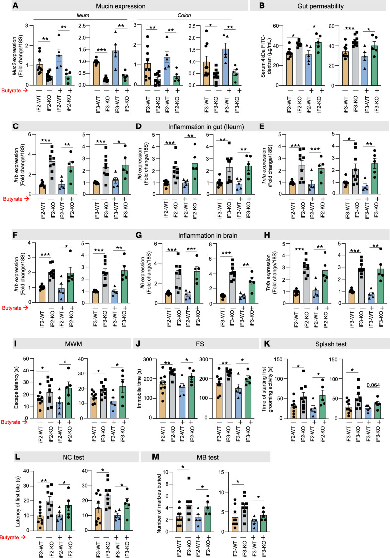
Aging-related abnormalities in gut microbiota are associated with cognitive decline, depression, and anxiety, but underlying mechanisms remain unstudied. Here, our study demonstrated that transplanting old gut microbiota to young mice induced inflammation in the gut and brain coupled with cognitive decline, depression, and anxiety. We observed diminished mucin formation and increased gut permeability ("leaky gut") with a reduction in beneficial metabolites like butyrate because of decline in butyrate-producing bacteria in the aged gut microbiota. This led to suppressed expression of butyrate receptors, free fatty acid receptors 2 and 3 (FFAR2/3). Administering butyrate alleviated inflammation, restored mucin expression and gut barriers, and corrected brain dysfunction. Furthermore, young mice with intestine-specific loss of FFAR2/3 exhibited gut and brain abnormalities akin to those in older mice. Our results demonstrate that reduced butyrate-producing bacteria in aged gut microbiota result in low butyrate levels and reduced FFAR2/3 signaling, leading to suppressed mucin formation that increases gut permeability, inflammation, and brain abnormalities. These findings underscore the significance of butyrate-FFAR2/3 agonism as a potential strategy to mitigate aged gut microbiota-induced detrimental effects on gut and brain health in older adults.

Keywords: Aging; Alzheimer disease; Bioinformatics; G protein–coupled receptors; Microbiology.


Conflict of interest statement

Conflict of interest: HY is a cofounder and chief scientific officer of Postbiotics Inc.

Figures

Figure 1
Figure 1. Old mouse gut microbiota is significantly distinct from that of sex-matched controls.
(A) Principal component analysis (PCA) of β-diversity shows that the microbiota composition significantly differs between old and young feces. Microbiome β-diversity was assessed using the Bray-Curtis dissimilarity index and visualized with PCA. (B and C) The abundance of major phyla and genera also differs between young and old microbiomes. (D) The cladogram of major genera levels in old versus young fecal microbiomes. (EH) Old (donor) mice show an increase in gut permeability (FITC-dextran leakage from gut to blood) (E); expression of inflammatory genes like Il1b, Il6, and Tnfa (F) in the intestine (ileum and colon); and higher circulating IL-6 (G) and TNF-α (H) compared with young controls. All values represent the mean of 5–10 animals in each group, and error bars represent the standard error of means. Statistical significance was determined using t tests (2 tailed) and/or ANOVA (1 or 2 way, as appropriate), and **P < 0.01 and ***P < 0.001 indicate statistical significance.
Figure 2
Figure 2. Old gut microbiota transplantation induces age-related leaky gut, inflammation, and behavioral abnormalities, and young microbiota transplantation reduces them.
(AC) Old FMT significantly increased leaky gut (FITC-dextran leakage) (A) and inflammatory markers (Il1b, Il6, and Tnfa) in the ileum (B) and brain (C) of recipient mice. (DI) Old FMT instigated behavioral changes, such as cognitive dysfunction (Morris water maze [MWM] test) (D), depression (forced swim [FS] and splash tests) (E and F), and anxiety (novel cage [NC], marble burying [MB], and open field [OF] tests) (GI). (JR) Interestingly, young FMT to old mice significantly ameliorated leaky gut (J), inflammation in the ileum (K) and brain (L), and cognitive decline (M), depression (N and O), and anxiety (PR) behaviors. All values represent the mean of 5–10 animals in each group, and error bars represent the standard error of the means. Statistical significance was determined using a 2-tailed t test, as applicable, and P values of *P < 0.05, **P < 0.01, and ***P < 0.001 are statistically significant.
Figure 3
Figure 3. Old microbiota disrupts gut barrier functions by reducing mucin expression and goblet cells in recipient mouse gut.
(A) Differential abundance data on an intestinal epithelia-related gene array showing significantly reduced expression of mucin genes Muc2 and Muc6 and tight junction gene Zo1 and increased expression of inflammatory genes Il6 and Tnfa in the intestine (ileum) of mice receiving FMT from old mice compared with controls receiving young FMT. (B) Random forest analysis shows that Muc2 was most significantly affected by old FMT. The mean decrease in gini score respresents the importance of the variable in building the model; thus, the higher the value of the mean decrease in gini score, the higher the importance of the variable in the model. (C and D) Old FMT recipients have significantly fewer goblet cells (periodic acid–Schiff [PAS] staining; red arrows) in their intestinal villi than controls. (E) Fecal mucin content was also significantly lower in old FMT recipients than in controls. (F and G) Treatment with fecal conditioned media (FCM) made from the feces of old mice significantly reduced transepithelial electrical resistance (TEER) (F) and increased FITC-dextran permeability (G) in the monolayers of human HT29 cells compared with treatment with young FCM. (HK) Muc2 and Muc6 expression was significantly reduced in goblet-like CMT93 cells (H) and enteroids (I) treated with old FCM; they resembled the intestines (ileum and colon) of old FMT recipient mice (J and K). All the values represent the mean of 5–10 animals or 3–4 independent replicates for each group in the cell and enteroid experiments repeated 2–3 times, and error bars represent the standard error of means. Statistical significance was determined using t test and/or ANOVA, as applicable, and P values *P < 0.05, **P < 0.01, and ***P < 0.001 are statistically significant.
Figure 4
Figure 4. Deficiency of butyrate-producing bacteria lowers butyrate levels in older gut.
(A) Principal component analysis (PCA) shows a significantly distinct metabolomics signature in the feces of old mice compared with that of sex-, diet-, and genotype-matched young mice. (B and C) Volcano plot (B) and random forest analyses (C) of differential abundance show that butyrate was the most significantly reduced metabolite in the old feces. Red dots represent metabolites more than +1 or –1 log2 fold-change whereas blue represents statistical significance but log2 fold-change is less than –1 or +1. (D and E) Compared to young feces, old feces showed significantly less expression of the butyrate-producing bacteria abundance markers butyrate kinase (buk) (D) and butyryl-CoA:acetate CoA transferase (but) (E) genes. (F) FMT transferred the reduced butyrate phenotype from old gut. (G and H) Reduced expression of buk (G) and but (H) was also transferred by old FMT. All the values represent the mean of 5–10 samples in each group. Statistical significance was determined using t test, and P values *P < 0.05 and ***P < 0.001 are statistically significant.
Figure 5
Figure 5. Butyrate treatment promotes gut mucin, protecting against the adverse effects of old microbiota on leaky gut and inflammation.
(AC) Old FCM reduced mucin accumulation (PAS [blue] staining) (A) and Muc2 expression in both CMT93 cells (B) and enteroids (C), and butyrate treatment (6 mM) attenuated the decrease. The original magnification of these images was 4×. (DF) Butyrate (2%) feeding in drinking water significantly reduced leaky gut (FITC-dextran leakage from gut to blood) (D) and increased Muc2 expression in the ileum (E) and fecal mucin content (F) of mice receiving old FMT (green) compared with controls not treated with butyrate (grey). Their gut recovered to the point that it resembled that of the young FMT recipient controls (blue). (GM) Similarly, old FMT-induced inflammation (Il1b, Il6, and Tnfa) in ileum (G) and brain (H) along with behavioral abnormalities, such as cognitive dysfunction (I), depression (J and K), and anxiety (L and M), were significantly reduced in butyrate-treated old FMT recipient mice (green) compared with controls (gray), until their condition resembled that of young FMT recipients (blue). All the values represent the mean of 5–8 animals or 3–4 independent replicates for each group in the cell and enteroid experiments, repeated 2–3 times, and error bars represent the standard error of means. Statistical significance was determined using 2-tailed Student’s t test or 1- or 2-way ANOVA, as applicable, and P values *,#P < 0.05, **P < 0.01, and ***,###P < 0.001 are statistically significant.
Figure 6
Figure 6. Old microbiota transplantation reduces FFAR2/3 signaling, which attenuates butyrate’s beneficial effect on gut mucin.
(A) Mice that received old FMT expressed significantly fewer Ffar2 and Ffar3 genes than those that received young FMT. (BD) Ffar2/3 expression was also significantly reduced in enteroids (B), CMT93 cells (C), and HT29 cells (D) treated with old FCM compared with controls treated with young FCM. (E) Changes recapitulated the suppressed Ffar2/3 expression in the intestine (ileum) of old donors compared with young controls (orange). (F and G) Butyrate (6 μM) significantly increases Ffar2/3 expression in non-FCM and young FCM-treated CMT93 cells and protects from decline in Ffar2/3 expression old FCM-treated cells, while such changes are not seen in acetate-treated groups. (H and I) Inhibiting FFAR2 (using CATPB) and FFAR3 (using siRNA) dampened the positive effects of butyrate treatment on mucin accumulation (PAS staining) (H) and Muc2, Muc6, and Muc13 expression (I) in CMT93 cells, indicating that FFAR2/3 signaling mediates butyrate’s increasing mucin. The original magnification for these images was 4×. All the values represent the mean of 5–10 animals or 3–4 independent replicates from each group in cells and enteroid experiments, repeated 2–3 times, and error bars represent the standard error of means. Statistical significance was determined using t test and/or ANOVA, as applicable, and P values *P < 0.05, **P < 0.01, and ***P < 0.001 are statistically significant.
Figure 7
Figure 7. FFAR2/3 deficiency in the gut exacerbates early aging in the brain, including neuroinflammation and behavioral changes.
(A) The expression of Muc2 in both ileum and colon was significantly lower in the 7-month-old, intestine-specific FFAR2 (iF2) and FFAR3 (iF3) knockout (KO) mice compared with their age- and sex-matched wild-type (WT) controls. (BE) In line, the gut permeability (FITC-dextran leakage) (B) was higher and the expression of inflammatory genes (Il1b, Il6, and Tnfa) (CE) was lower in the intestine of the 7-month-old iF2/3 KO mice than in WT controls. (FM) The brains of the 7-month-old iF2/3-KO mice showed significantly higher levels of inflammatory markers (Il1b, Il6, and Tnfa) (FH) along with cognitive decline (MWM test) (I), depression (FS and splash tests) (J and K), and anxiety-like behaviors (NC and MB tests) (L and M) compared with WT controls. (AM) No significant changes in Muc2 expression (A), gut permeability (B), inflammation in the gut (CE) and brain (FH), and behavioral changes (IM) were seen in iF2/3-KO mice compared to WT controls. All the values represent the mean of 5–10 animals in each group, and error bars represent the standard error of means. Statistical significance was determined using ANOVA and/or t test, and P values *P < 0.05, **P < 0.01, and ***P < 0.001 are statistically significant.

References

    1. Cullen NC, et al. Accelerated inflammatory aging in Alzheimer’s disease and its relation to amyloid, tau, and cognition. Sci Rep. 2021;11(1):1965. doi: 10.1038/s41598-021-81705-7. - DOI - PMC - PubMed
    1. Gomez-Nicola D, Boche D. Post-mortem analysis of neuroinflammatory changes in human Alzheimer’s disease. Alzheimers Res Ther. 2015;7(1):42. doi: 10.1186/s13195-015-0126-1. - DOI - PMC - PubMed
    1. Kennedy BK, et al. Geroscience: linking aging to chronic disease. Cell. 2014;159(4):709–713. doi: 10.1016/j.cell.2014.10.039. - DOI - PMC - PubMed
    1. Ferrucci L, Fabbri E. Inflammageing: chronic inflammation in ageing, cardiovascular disease, and frailty. Nat Rev Cardiol. 2018;15(9):505–522. doi: 10.1038/s41569-018-0064-2. - DOI - PMC - PubMed
    1. Koelman L, et al. Cytokines for evaluation of chronic inflammatory status in ageing research: reliability and phenotypic characterisation. Immun Ageing. 2019;16:11. doi: 10.1186/s12979-019-0151-1. - DOI - PMC - PubMed

Publication types