Review
. 2024 Jul 1;104(3):1121-1145.
doi: 10.1152/physrev.00019.2023. Epub 2024 Feb 8.

The hormonal control of parturition

Affiliations
Review

The hormonal control of parturition

Emily Hamburg-Shields et al. Physiol Rev. .

Abstract

Parturition is a complex physiological process that must occur in a reliable manner and at an appropriate gestation stage to ensure a healthy newborn and mother. To this end, hormones that affect the function of the gravid uterus, especially progesterone (P4), 17β-estradiol (E2), oxytocin (OT), and prostaglandins (PGs), play pivotal roles. P4 via the nuclear P4 receptor (PR) promotes uterine quiescence and for most of pregnancy exerts a dominant block to labor. Loss of the P4 block to parturition in association with a gain in prolabor actions of E2 are key transitions in the hormonal cascade leading to parturition. P4 withdrawal can occur through various mechanisms depending on species and physiological context. Parturition in most species involves inflammation within the uterine tissues and especially at the maternal-fetal interface. Local PGs and other inflammatory mediators may initiate parturition by inducing P4 withdrawal. Withdrawal of the P4 block is coordinated with increased E2 actions to enhance uterotonic signals mediated by OT and PGs to promote uterine contractions, cervix softening, and membrane rupture, i.e., labor. This review examines recent advances in research to understand the hormonal control of parturition, with focus on the roles of P4, E2, PGs, OT, inflammatory cytokines, and placental peptide hormones together with evolutionary biology of and implications for clinical management of human parturition.

Keywords: hormonal; human; parturition; pregnancy.


Conflict of interest statement

No conflicts of interest, financial or otherwise, are declared by the authors.

Figures

None
Graphical abstract
FIGURE 1.
FIGURE 1.
Endocrine, paracrine, and neuroendocrine interactions controlling parturition. Multiple physiological factors (some examples shown) contribute signals impacting uterine tissues that are integrated into a putative inflammatory load. In a threshold-limited manner, inflammatory load induces progesterone (P4)/P4 receptor (PR) withdrawal leading to loss of the P4/PR block to labor, which includes increased local inflammation due to loss of P4/PR anti-inflammatory activity, leading to increased local production of inflammatory cytokines, some of which [e.g., prostaglandin (PG)F] are potent uterotonins. P4/PR withdrawal also increases expression of ESR1 [encodes estrogen receptor-α (ERα)], allowing circulating estrogens (mainly estradiol) to increase response of uterine tissues to uterotonins [especially PGF and oxytocin (OT)] and increase extracellular matrix (ECM) breakdown in the cervix and fetal membrane, leading to cervix softening and membrane weakening, respectively. A neuroendocrine positive feedback loop involving OT amplifies contractions at the time of active labor: phasic myometrium contractions, cervix softening and dilation, and membrane rupture.
FIGURE 2.
FIGURE 2.
The majority of pregnancy is a quiescent state, with low myometrial contractile activity, maintained by progesterone (P4), which is the dominant hormonal effector of ongoing gestation. The progression from quiescent pregnancy to latent (early) labor is facilitated by a functional progesterone withdrawal and increased estradiol (E2) effect. Uterotonins [prostaglandins (PGs) and oxytocin (OT)] are the predominant effectors of active labor and postpartum uterine involution. Image is an adaptation of material from Ref. .
FIGURE 3.
FIGURE 3.
Signaling pathways by which progesterone (P4)/P4 receptor (PR) blocks labor. P4/PR via decidual cell expression of IL-15 promotes immune quiescence at the maternal-fetal interface. P4/PR directly inhibits expression of ESR1, which decreases levels of estrogen receptor-α (ERα), leading to refractoriness to estradiol (E2)/ERα-induced expression of contraction-associated protein (CAP) genes. P4/PR increases expression of ZEB1, whose downstream effects inhibit CAP and inflammatory cytokine gene expression. P4/PR interacts physically with the activator protein-1 (AP-1) transcription factor complex to inhibit activity of AP-1 and NF-κB, leading to a broad anti-inflammatory effect and inhibition of AP-1-driven CAP genes. For most of pregnancy P4/PR effects (green) dominate. STAT5b, signal transducer and activator of transcription 5b; 20αHSD, 20α-hydroxysteroid dehydrogenase.
FIGURE 4.
FIGURE 4.
Modes of progesterone (P4) withdrawal. Systemic P4 withdrawal occurs by P4 metabolism in the source tissue [e.g., corpus luteum (CL) via 20α-hydroxysteroid dehydrogenase (20αHSD) or placenta via P450c17]. Similarly, local withdrawal occurs by P4 metabolism by 20αHSD in uterine target cells to prevent P4 from interacting with P4 receptors (PRs). Functional P4 withdrawal occurs by changes in PR transcriptional activity caused by interaction with specific repressors, transrepression by phosphorylated PR-A, and/or direct activation of contraction-associated protein (CAP) gene expression by liganded PR-A. A4, androstenedione; ERα, estrogen receptor-α; 20αOHP, 20α-hydroxyprogesterone.
FIGURE 5.
FIGURE 5.
Comparison of endocrine and paracrine pathways to progesterone (P4)/P4 receptor (PR) withdrawal in human (solid line), ovine (dashed line), and murine (dotted line) pregnancy. A comparative approach highlights the unique roles of the murine corpus luteum and ovine placental P4 metabolism in P4/PR withdrawal versus the suspected mechanism of functional P4 withdrawal in human myometrium/decidua. CRH, corticotropin-releasing hormone; DHEA, dehydroepiandrosterone; E2, estradiol; PGF, prostaglandin F. Image is an adaptation of material from Ref. .
FIGURE 6.
FIGURE 6.
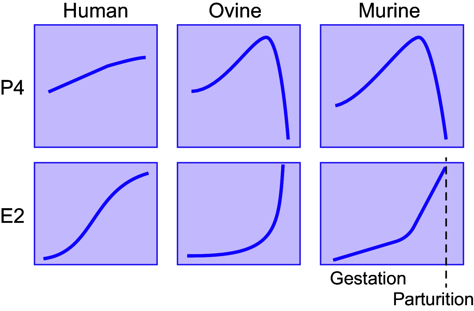
Comparison of relative serum levels of progesterone (P4) and estradiol (E2) throughout gestation in human, sheep, and mouse. The level of P4 drops precipitously immediately before parturition in sheep and mouse but in human remains persistently elevated until after parturition. A functional withdrawal of P4/P4 receptor (PR), therefore, is not reflected in serum hormone levels in the context of human pregnancy. Image is an adaptation of material from Ref. .
FIGURE 7.
FIGURE 7.
The inflammatory threshold hypothesis for the control of parturition timing. During pregnancy, inflammatory stimuli derived from multiple intrinsic (e.g., fetal development, placenta senescence) and extrinsic (e.g., maternal stress, intrauterine infection) factors contribute to a net inflammatory load impacting the uterine tissues. For most of pregnancy the progesterone (P4)/P4 receptor (PR) anti-inflammatory block to labor prevents uterine tissue-level inflammation. Above a threshold level, inflammatory stimuli induce P4 withdrawal via mechanism outlined in FIGURE 4. Loss of the P4/PR block to labor allows a positive-feedback proinflammatory state in the uterine tissues that induces active labor via increased contraction-associated protein (CAP) gene expression and increased production of and sensitivity to prolabor uterotonins. The timing of parturition is determined by the inflammatory load trajectory and the inflammatory load threshold for inflammation-induced P4/PR withdrawal. The threshold is normally reached at term but can be reached earlier in pathological states (e.g., intrauterine infection).
FIGURE 8.
FIGURE 8.
Proposed model in which maternal and fetal cortisol stimulate corticotropin-releasing hormone (CRH) production by the trophoblast. This would generate positive feedback loops to result in increased estradiol (E2) to promote maternal expression of contraction-associated proteins (CAPs) and contribute to the onset of parturition. Cortisol-induced fetal organ maturation is also positively affected by placental-fetal feedback in this system. DHEA, dehydroepiandrosterone. Image is an adaptation of material from Ref. .
FIGURE 9.
FIGURE 9.
Visual comparison of the relative proportions of the fetal skull to the maternal pelvis in primates shows a maximal cephalopelvic ratio in humans. The obstetric dilemma hypothesis proposes that the biomechanical limitations of the pelvic outlet (blue outline) and the size of the human head (red outline) necessitate that parturition occur at a time that results in relative altriciality of the human neonate. Image is an adaptation of material from Ref. .
FIGURE 10.
FIGURE 10.
The hormonal regulation of parturition provides an opportunity for therapeutic intervention, both for prevention of preterm labor (left) and for induction of labor (right). Interventions that directly target hormonal pathways are listed for the prevention of preterm labor (left) and induction of labor (right). Attempts to prevent preterm labor have targeted progesterone (P4) receptor (PR), oxytocin receptor (OTR), prostaglandin (PG) synthesis, and estrogen receptor (ER). These interventions include some with therapeutic benefit in a limited population (e.g., vaginal P4 for short cervix), some with unclear or unproven benefit (e.g., atosiban), and some harmful and contraindicated in pregnancy [e.g., diethylstilbestrol (DES)]. Medically indicated labor and management of miscarriage, in contrast, utilize all of the listed interventions routinely as the standard of care in common clinical scenarios.

References

    1. Mesiano S. The endocrinology of human pregnancy and fetoplacental neuroendocrine development. In: Yen and Jaffe's Reproductive Endocrinology: Physiology, Pathophysiology, and Clinical Management, edited by Strauss JF, Barbieri RL.. Philadelphia, PA: Elsevier, 2009, p. 249–282.
    1. Mesiano S, DeFranco E, Muglia L. Parturition. In: Knobil and Neill’s Physiology of Reproduction, edited by Plant TM, Zeleznik A.. London: Academic Press, 2014, p. 1857–1925.
    1. Casey ML, Macdonald PC. Endocrine changes of pregnancy. In: Williams Textbook of Endocrinology, edited by Wilson JD, Foster DW, Kronenberg HM, Larsen PR.. Philadelphia, PA: W.B. Saunders, 1998, p. 1259.
    1. Csapo A. The luteo-placental shift, the guardian of pre-natal life. Postgrad Med J 45: 57–64, 1969. doi: 10.1136/pgmj.45.519.57. - DOI - PMC - PubMed
    1. Csapo AI, Pulkkinen M. Indispensability of the human corpus luteum in the maintenance of early pregnancy. Lutectomy evidence. Obstet Gynecol Surv 33: 69–81, 1978. doi: 10.1097/00006254-197802000-00001. - DOI - PubMed

Publication types

LinkOut - more resources