Clinical Trial
. 2024 Jun;51(7):2085-2097.
doi: 10.1007/s00259-024-06630-z. Epub 2024 Feb 8.

Adjuvant holmium-166 radioembolization after radiofrequency ablation in early-stage hepatocellular carcinoma patients: a dose-finding study (HORA EST HCC trial)

Affiliations
Clinical Trial

Adjuvant holmium-166 radioembolization after radiofrequency ablation in early-stage hepatocellular carcinoma patients: a dose-finding study (HORA EST HCC trial)

Pim Hendriks et al. Eur J Nucl Med Mol Imaging. 2024 Jun.

Abstract

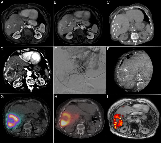
Purpose: The aim of this study was to investigate the biodistribution of (super-)selective trans-arterial radioembolization (TARE) with holmium-166 microspheres (166Ho-MS), when administered as adjuvant therapy after RFA of HCC 2-5 cm. The objective was to establish a treatment volume absorbed dose that results in an absorbed dose of ≥ 120 Gy on the hyperemic zone around the ablation necrosis (i.e., target volume).

Methods: In this multicenter, prospective dose-escalation study in BCLC early stage HCC patients with lesions 2-5 cm, RFA was followed by (super-)selective infusion of 166Ho-MS on day 5-10 after RFA. Dose distribution within the treatment volume was based on SPECT-CT. Cohorts of up to 10 patients were treated with an incremental dose (60 Gy, 90 Gy, 120 Gy) of 166Ho-MS to the treatment volume. The primary endpoint was to obtain a target volume dose of ≥ 120 Gy in 9/10 patients within a cohort.

Results: Twelve patients were treated (male 10; median age, 66.5 years (IQR, [64.3-71.7])) with a median tumor diameter of 2.7 cm (IQR, [2.1-4.0]). At a treatment volume absorbed dose of 90 Gy, the primary endpoint was met with a median absorbed target volume dose of 138 Gy (IQR, [127-145]). No local recurrences were found within 1-year follow-up.

Conclusion: Adjuvant (super-)selective infusion of 166Ho-MS after RFA for the treatment of HCC can be administered safely at a dose of 90 Gy to the treatment volume while reaching a dose of ≥ 120 Gy to the target volume and may be a favorable adjuvant therapy for HCC lesions 2-5 cm.

Trial registration: Clinicaltrials.gov NCT03437382 . (registered: 19-02-2018).

Keywords: Adjuvant therapy; Dose-escalation study; Hepatocellular carcinoma; Holmium-166; Radiofrequency ablation; Trans-arterial radioembolization.


Conflict of interest statement

M.C. Burgmans has received an educational grant from Boston Scientific and Medtronic and consultancy fees from SIRTeX, Medtronic, Delcath Systems, and Philips Health Care. None are related to the current project. J.F.W. Nijsen is co-founder of Quirem Medical, which has been acquired by Terumo Europe NV in July 2020. Nijsen has a scientific advisory role and is entitled to certain milestone payments from Terumo, which are related to Quirem’s financial, operational, and regulatory performance in the future. Furthermore, Nijsen is inventor on the patents related to radioactive microspheres that are assigned to University Medical Center Utrecht Holding BV, Quirem Medical, or BASF Corp. The activities of J.F.W. Nijsen within Quirem Medical are approved and supported by the Board of Directors of the Radboudumc. All other authors declare that they have no conflict of interest.

Figures

Fig. 1
Fig. 1
Schematic drawings of the study procedure. A HCC lesion of 2–5 cm. B Thermal ablation of HCC lesion. C Potential sites of local recurrences due to impaired heat propagation, heat-sink effect, or satellite nodules. D Target volume for adjuvant TARE. E Deposition of 166Ho-MS with preferential flow of microspheres to the hyperemic zone surrounding the ablated tissue (i.e., target volume). F Liver volume infused with 166Ho-MS TARE (i.e., treatment volume) [22].
Fig. 2
Fig. 2
Flowchart of the study population.
Fig. 3
Fig. 3
HORA EST HCC treatment sequence: A Arterial scan phase of diagnostic MRI showing a hypervascular HCC lesion of 31 mm in the liver. B Portal venous scan phase of MRI showing central wash-out in the HCC lesion. C Intraprocedural CT after placement of six cooled-tip RFA needles with 3-cm exposed tip. D Intraprocedural contrast enhanced CT scan in arterial phase showing hyperemia around the ablation zone on post-ablation CECT. E Super-selective catheterization of hepatic arteries with vascular supply to the target volume. F CBCT of the treatment volume with an identical catheter position as in E. G SPECT-CT of [99mTc]Tc-MAA dose distribution used for dose planning. H SPECT-CT of 166Ho-MS distribution. I MRI-based dosimetry of 166Ho-MS distribution.
Fig. 4
Fig. 4
Dose distribution per patient within treatment volume, based on SPECT imaging. The bars in black represent the mean absorbed dose on the target volume directly surrounding the ablation volume per patient. The cutoff point of an absorbed target volume dose of 120 Gy is indicated by the horizontal dashed line. The bars in white show the absorbed dose to the non-target volume within the treatment volume. The first two patients were treated with 60 Gy to the treatment volume, whereas the other patients were treated with 90 Gy to the treatment volume. The median ratio of target volume dose vs non-target volume dose was 1.97 (IQR, [1.75–2.17]).
Fig. 5
Fig. 5
Histology of explanted liver treated with radiofrequency ablation and adjuvant 166Ho TARE. Digitalized histology using Ultra Fast Scanner (Philips Healthcare, Best, The Netherlands) with a magnitude of 40×. A Zoom 10×. Transition from liver tissue with ductal proliferation to fibrosis with marked depositions of 166Ho-MS. B Zoom 5×. Overview of transition from ductal proliferation to necrotic tissue with marked 166Ho-MS.

References

    1. Galle PR, Forner A, Llovet JM, Mazzaferro V, Piscaglia F, Raoul J-L, et al. EASL Clinical Practice Guidelines: management of hepatocellular carcinoma. J Hepatol. 2018;69:182–236. doi: 10.1016/j.jhep.2018.03.019. - DOI - PubMed
    1. Reig M, Forner A, Rimola J, Ferrer-Fábrega J, Burrel M, Garcia-Criado Á, et al. BCLC strategy for prognosis prediction and treatment recommendation: The 2022 update. J Hepatol. 2022;76(3):681–693. doi: 10.1016/j.jhep.2021.11.018. - DOI - PMC - PubMed
    1. Forner A, Reig M, Bruix J. Hepatocellular carcinoma. Lancet. 2018;391:1301–1314. doi: 10.1016/S0140-6736(18)30010-2. - DOI - PubMed
    1. Weis S, Franke A, Mössner J, Jakobsen JC, Schoppmeyer K. Radiofrequency (thermal) ablation versus no intervention or other interventions for hepatocellular carcinoma. Cochrane Database Syst Rev. 2013;(12):Cd003046. 10.1002/14651858.CD003046.pub3. - PMC - PubMed
    1. Xu X-L, Liu X-D, Liang M, Luo B-M. Radiofrequency ablation versus hepatic resection for small hepatocellular carcinoma: systematic review of randomized controlled trials with meta-analysis and trial sequential analysis. Radiology. 2017;287:461–472. doi: 10.1148/radiol.2017162756. - DOI - PubMed

Associated data