. 2024 Apr 23;8(8):1869-1879.
doi: 10.1182/bloodadvances.2023012155.

Human Fc gamma receptor IIIA blockade inhibits platelet destruction in a humanized murine model of ITP

Affiliations

Human Fc gamma receptor IIIA blockade inhibits platelet destruction in a humanized murine model of ITP

Lazaro Gil Gonzalez et al. Blood Adv. .

Abstract

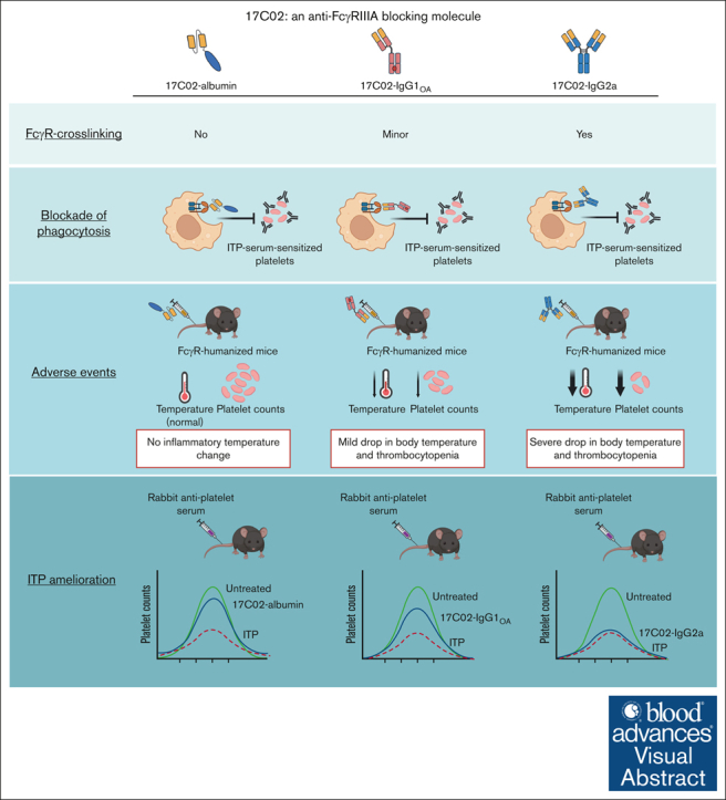
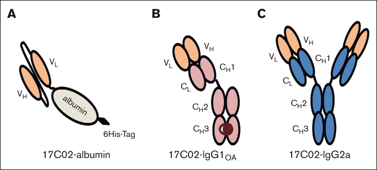
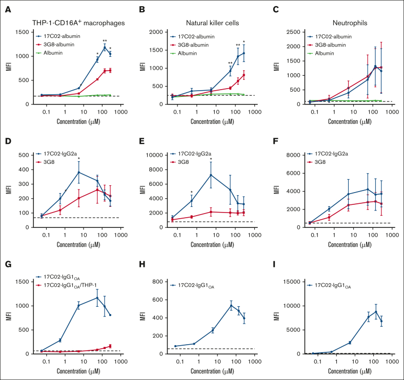
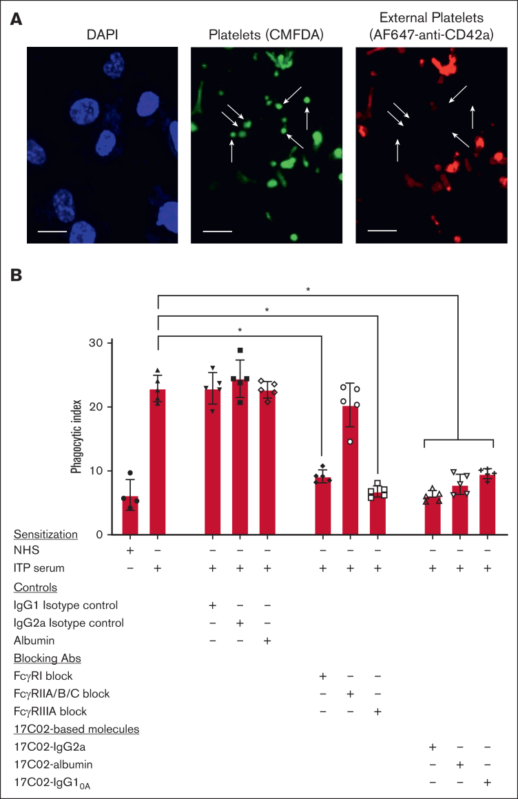
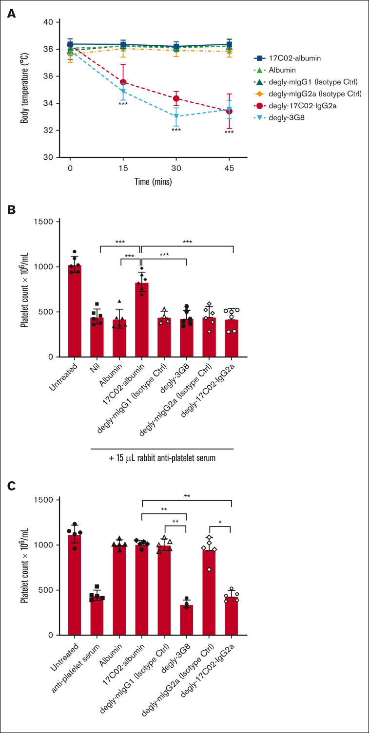
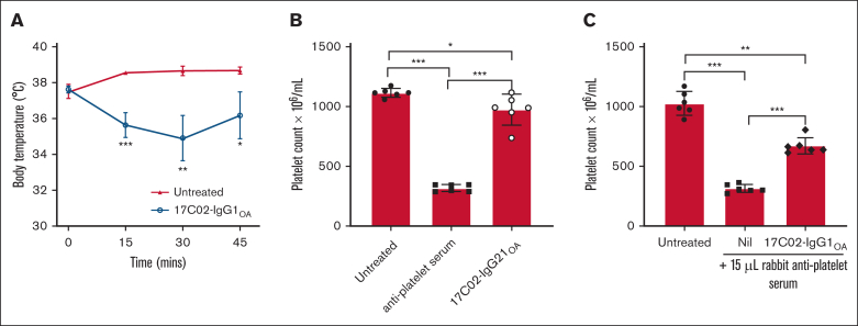
Fc gamma receptor (FcγR) IIIA is an important receptor for immunoglobulin G (IgG) and is involved in immune defense mechanisms as well as tissue destruction in some autoimmune diseases including immune thrombocytopenia (ITP). FcγRIIIA on macrophages can trigger phagocytosis of IgG-sensitized platelets, and prior pilot studies observed blockade of FcγRIIIA increased platelet counts in patients with ITP. Unfortunately, although blockade of FcγRIIIA in patients with ITP increased platelet counts, its engagement by the blocking antibody drove serious adverse inflammatory reactions. These adverse events were postulated to originate from the antibody's Fc and/or bivalent nature. The blockade of human FcγRIIIA in vivo with a monovalent construct lacking an active Fc region has not yet been achieved. To effectively block FcγRIIIA in vivo, we developed a high affinity monovalent single-chain variable fragment (scFv) that can bind and block human FcγRIIIA. This scFv (17C02) was expressed in 3 formats: a monovalent fusion protein with albumin, a 1-armed human IgG1 antibody, and a standard bivalent mouse (IgG2a) antibody. Both monovalent formats were effective in preventing phagocytosis of ITP serum-sensitized human platelets. In vivo studies using FcγR-humanized mice demonstrated that both monovalent therapeutics were also able to increase platelet counts. The monovalent albumin fusion protein did not have adverse event activity as assessed by changes in body temperature, whereas the 1-armed antibody induced some changes in body temperature even though the Fc region function was impaired by the Leu234Ala and Leu235Ala mutations. These data demonstrate that monovalent blockade of human FcγRIIIA in vivo can potentially be a therapeutic strategy for patients with ITP.


Conflict of interest statement

Conflict-of-interest disclosure: Patent applications describing the 17C02-based molecules are assigned to the Canadian Blood Services with the participation of adMare BioInnovations and Unity Health/St. Michael’s Hospital. A.H.L. has had research funding from CSL and has other patents on IV immunoglobulin alternatives. The remaining authors declare no competing financial interests.

Figures

None
Graphical abstract
Figure 1.
Figure 1.
Schematic representation for obtaining scFvs that bind and block FcγRIIIA. BALB/c mice were immunized with the recombinant human FcγRIIIA, total splenic RNA was isolated, and genes encoding the VH and VL chains were amplified. A second polymerase chain reaction stitched VH and VL with a linker, and the products were cloned into a phagemid vector via Gibson assembly. E coli was then transformed with the constructs and a scFv-phage display library obtained. Five rounds of selection (R1, R2A, R2B, R3A, and R3B) were performed to select phages bound to FcγRIIIA with minimal cross-reactivity with FcγRIIA (supplemental Figure 1). Selection of scFv was based on binding to NK cells by flow cytometry and inhibition of hIgG-FcγRIIIA interaction by homogeneous time-resolved fluorescence. Purified scFv were analyzed for binding to FcγRIIIA by ELISA and Octet; minimal cross-reactivity with the other human receptors and inhibition of hIgG-FcγRIIIA interaction (ie, FcγRIIIA blockers) were part of the selection process. The final antibody fragment, 17C02-scFv, was selected from 10 candidates based on these assessments as well as sequencing analysis to screen for glycosylation, oxidation, aggregation, deamidation/isomerization, and proteolytic sites to exclude scFv molecules with low biochemical stability.
Figure 2.
Figure 2.
Schematic representation of the 17C02-based molecules. (A) The gene that encodes for the 17C02-scFv with a linker (connecting the VH with the VL chains) as well as an additional RGGGGSGGGGS were used to connect the scFv to the N-terminal sequence of human albumin and a 6xHis tag at the C-terminal end of albumin. (B) The genes encoding the VL and VH of 17C02 were used to express a human IgG1 1-armed antibody with the LALA mutation to impair Fc-FcγR interactions, using the “knob-into-hole” strategy. (C) The genes that encode for the VL and VH of 17C02 were expressed as a full-length mouse IgG2a antibody.
Figure 3.
Figure 3.
Binding of 17C02-based molecules to human FcγRIIIA expressed on THP-1-CD16A cells, THP-1 cells, as well as primary cells. Cells were incubated with the indicated concentration of either 17C02-based molecules, 3G8-based molecules, or albumin alone (negative control). The following were the cell types used for each panel: (A,D) THP-1-CD16A cells; (B,E,H) NK cells from healthy human donors; and (C,F,I) neutrophils from healthy human donors; as well as (G) a comparison between THP-1-CD16A and THP-1 cells. Binding of albumin fusion proteins was detected using a FITC-labeled monoclonal antihuman albumin antibody, whereas binding of deglycosylated full-length antibodies or the 1-armed antibody was detected using an APC-labeled goat F(ab’)2 antimouse IgG (Fc-specific) or an AF647-labeled donkey antihuman IgG (H + L), respectively. Stained cells were washed and analyzed by flow cytometry using the BD LSRFortessa X-20. Data analysis was performed using FlowJo v10. Data are presented as mean ± standard deviation from 3 to 5 independent experiments. The dashed line represents the mean fluorescent intensity (MFI; arbitrary units) value for the corresponding secondary antibody alone at 5 μg/mL. Statistical analysis was performed using a 2-way analysis of variance (ANOVA) and Sidak multiple comparisons test, comparing the MFI values of the 3 molecules at each antibody concentration (∗P < .05; ∗∗P < .01).
Figure 4.
Figure 4.
Blocking capacity of 17C02-based molecules and FcγR utilization by THP-1-CD16A cells in the phagocytosis of IgG-opsonized human platelets. (A) Images of platelets sensitized with ITP serum and later incubated with THP-1-CD16A macrophages. Images were taken at the center of each well with Z-stacking. Phagocytosis was quantified using Imaris v9.6.0. The white arrows denote examples of phagocytosis of platelets; scale bar, 10 μm. (B) PI from 4 independent experiment are shown. Sensitization: “+” indicates platelets were incubated with normal human serum vs serum from patients with ITP. The PI was calculated as the number of platelets engulfed per 100 macrophages. The contribution of FcγRI, II, and III to phagocytosis was evaluated using Fc region deglycosylated blocking antibodies (final concentration of 10 μg/mL; 0.07 μM each): anti-FcγRI (clone 10.1), anti-FcγRIIA/B/C (clone AT10), or anti-FcγRIIIA (clone 3G8). The deglycosylated mouse IgG1 (clone MOPC-21), the deglycosylated mouse IgG2a (clone N/A-CP150), and human albumin were used as controls (final concentration of 0.07 μM). The blocking capacity of 17C02-based molecules was evaluated (17C02-albumin, 17C02-IgG1OA, and deglycosylated 17C02-IgG2a) using the same comparative final molar concentration. Data are presented as the mean ± the standard deviation (n = 4-5). The statistical analysis was performed using Kruskal-Wallis and Dunn multiple comparison test (∗P < .05).
Figure 5.
Figure 5.
Ameliorative effects and adverse events caused by 17C02-based molecules and 3G8 in an antibody-mediated model of ITP. FcγR-humanized mice were treated with an IV administration of either deglycosylated full-length 17C02 or deglycosylated full-length 3G8 (81 μg/mouse; 540 μM/mouse), 17C02-albumin (50 μg/mouse; 540 μM/mouse), deglycosylated full-length IgG1 and IgG2 isotype controls (81 μg/mouse; 540 μM/mouse), or albumin alone (35.1 μg/mouse; 540 μM/mouse). (A) Decreases in rectal temperature were evaluated as an indicator of an inflammatory adverse event comparing 0-minute (pretreatment) with 15-, 30-, and 45-minute posttreatment conditions. Data are presented as mean ± standard deviation (n = 5-7). The statistical analysis was performed by a 2-way ANOVA and Sidak multiple comparisons test (∗∗∗P < .001). (B) ITP was then induced in mice with 15 μL of a rabbit antiplatelet serum 2 hours after the anti-FcγRIIIA therapeutic intervention. Additional mice were either left untreated (Untreated) or treated with the antiplatelet serum alone (Nil) as comparative controls. Data are presented as mean ± standard deviation (n = 6). The statistical analysis was performed by a 1-way ANOVA and Tukey multiple comparisons test (∗∗∗P < .001). (C) The ability of FcγRIIIA-blocking reagents and controls to directly induce thrombocytopenia (as an adverse event) was evaluated 2 hours after treatment. The antiplatelet serum alone (15 μL/mouse) was used as a positive control. Deglycosylated mouse IgG1 (degly-mIgG1) and IgG2a (degly-mIgG2a) isotype controls were used as negative controls for amelioration. Data are presented as mean ± standard deviation (n = 5). The statistical analysis was performed using Kruskal-Wallis and Dunn multiple comparison test (∗P < .05; ∗∗P < .01).
Figure 6.
Figure 6.
Ameliorative effects and adverse events caused by 17C02-IgG1OA in an antibody-mediated model of ITP. (A) FcγR-humanized mice were IV injected with 540 μM of 17C02-IgG1OA, and body temperatures of mice were assessed for 45 minutes after treatment to investigate the inflammatory nature of the molecule (time “0” indicates before treatment). Data are presented as mean ± standard deviation (n = 6). The statistical analysis was performed by a 2-way ANOVA and Sidak multiple comparisons test (∗P < .05; ∗∗P < .01; ∗∗∗P < .001). (B) Mice were bled and platelet counts assessed 2 hours after 17C02-IgG1OA treatment to determine the ability of the antibody itself to cause thrombocytopenia. Rabbit antiplatelet serum alone (15 μL/mouse) was used as a positive control. Data are presented as mean ± standard deviation from 3 independent experiments (n = 6). The statistical analysis was performed by a 1-way ANOVA and Tukey multiple comparisons test (∗P < .05; ∗∗∗P < .001). (C) Mice were IV injected with 15 μL of rabbit antiplatelet serum 2 hours after 17C02-IgG1OA treatment to induce thrombocytopenia. Two hours after injection with the antiplatelet serum, mice were bled for enumeration of platelet counts to assess the ability of 17C02-IgG1OAto ameliorate thrombocytopenia. Data are presented as mean ± standard deviation (n = 6 mice). The statistical analysis was performed by a 1-way ANOVA and Tukey multiple comparisons test (∗∗P < .01; ∗∗∗P < .001).

References

    1. Olingy CE, Dinh HQ, Hedrick CC. Monocyte heterogeneity and functions in cancer. J Leukoc Biol. 2019;106(2):309–322. - PMC - PubMed
    1. Semple JW, Rebetz J, Maouia A, Kapur R. An update on the pathophysiology of immune thrombocytopenia. Curr Opin Hematol. 2020;27(6):423–429. - PubMed
    1. Audia S, Grienay N, Mounier M, Michel M, Bonnotte B. Evans’ syndrome: from diagnosis to treatment. J Clin Med. 2020;9(12):3851. - PMC - PubMed
    1. Kurlander RJ, Ellison DM, Hall J. The blockade of Fc receptor-mediated clearance of immune complexes in vivo by a monoclonal antibody (2.4G2) directed against Fc receptors on murine leukocytes. J Immunol. 1984;133(2):855–862. - PubMed
    1. Clarkson SB, Bussel JB, Kimberly RP, Valinsky JE, Nachman RL, Unkeless JC. Treatment of refractory immune thrombocytopenic purpura with an anti-Fc gamma-receptor antibody. N Engl J Med. 1986;314(19):1236–1239. - PubMed

Publication types