. 2024 Feb 15:100:117613.
doi: 10.1016/j.bmc.2024.117613. Epub 2024 Jan 28.

Discovery of small molecule benzothiazole and indole derivatives tackling tau 2N4R and α-synuclein fibrils

Affiliations

Discovery of small molecule benzothiazole and indole derivatives tackling tau 2N4R and α-synuclein fibrils

Ahmed A Elbatrawy et al. Bioorg Med Chem. .

Abstract

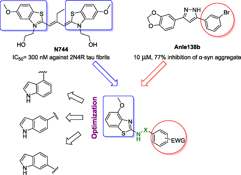
Tau and α-synuclein aggregates are the main histopathological hallmarks present in Alzheimer's disease (AD), Parkinson's disease (PD), and other neurodegenerative disorders. Intraneuronal hyperphosphorylated tau accumulation is significantly connected to the degree of cognitive impairment in AD patients. In particular, the longest 2N4R tau isoform has a propensity to rapidly form oligomers and mature fibrils. On the other hand, misfolding of α-synuclein (α-syn) is the characteristic feature in PD and dementia with Lewy bodies (DLB). There is a strong crosstalk between the two prone-to-aggregation proteins as they coprecipitated in some brains of AD, PD, and DLB patients. Simultaneous targeting of both proteinaceous oligomers and aggregates is still challenging. Here, we rationally designed and synthesized benzothiazole- and indole-based compounds using the structural hybridization strategy between the benzothiazole N744 cyanine dye and the diphenyl pyrazole Anle138b that showed anti-aggregation activity towards 2N4R tau and α-syn, respectively. The anti-aggregation effect of the prepared compounds was monitored using the thioflavin-T (ThT) fluorescence assay, while transmission electron microscopy (TEM) was employed to detect fibrils upon the completion of a time-course study with the ThT assay. Moreover, the photo-induced crosslinking of unmodified protein (PICUP) assay was used to determine the formation of oligomers. Specifically, compounds 46 and 48 demonstrated the highest anti-aggregation activity by decreasing the ThT fluorescence to 4.0 and 14.8%, respectively, against α-syn. Although no noticeable effect on 2N4R tau oligomers, 46 showed promising anti-oligomer activity against α-syn. Both compounds induced a significantly high anti-aggregation effect against the two protein fibrils as visualized by TEM. Moreover, compound 48 remarkably inhibited α-syn inclusion and cell confluence using M17D cells. Collectively, compounds 46 and 48 could serve as a basic structure for further optimization to develop clinically active AD and PD disease-modifying agents.

Keywords: Alpha-synuclein; Alzheimer’s disease; Anti-oligomer agents; Parkinson’s disease; Tau isoforms.


Conflict of interest statement

Declaration of competing interest The authors declare that they have no known competing financial interests or personal relationships that could have appeared to influence the work reported in this paper.

Figures

Fig. 1.
Fig. 1.
Design of methoxy benzothiazole and indole derivatives as dual 2N4R tau and α-syn aggregates inhibitors.
Fig. 2.
Fig. 2.
ThT fluorescence assay. The ThT kinetic curves of compounds 46, 47, and 48 (100 μM) when tested with: A. α-syn (2 μM); B. Dose-dependent inhibition of varying concentrations (3.125, 6.25, 12.5, 25, 50, 100 μM) of compound 46 on α-syn (2 μM) fibril formation using ThT fluorescence assay. For each concentration, triplicate data were gathered at the plateau phase from five consecutive time points. The error bars represent the individual standard error of the mean (SEM) for each condition.
Fig. 3.
Fig. 3.
Inhibition of the α-syn oligomer formation induced by the PICUP assay. In the PICUP experiment, α-syn (60 μM) was cross-linked with varying concentrations of compounds 46, 47, and 48 (50, 100, and 200 μM). The optical density of the high molecular-weight bands represents α-syn oligomer while the lower bands represent the protein monomers. In particular, compound 46 reduced the development of α-syn oligomer-corresponding high molecular bands between 35 and 40 kDa. The control consisted of no light exposure and no Tris(2,2′-bipyridyl)ruthenium(II) chloride (Ru(bpy), a cross-linking agent).
Fig. 4.
Fig. 4.
Inhibition of the oligomer formation induced by the PICUP assay on two different tau isoforms (10 μM): A. 2N4R tau; B. 0N4R tau. In the experiment, we exposed the protein to different concentrations of compounds 46, 47, and 48 (50 μM, 100 μM, and 200 μM). None of the compounds demonstrated the ability to suppress the formation of tau oligomers using both tau isoforms. The control consisted of no light exposure and no Tris(2,2′-bipyridyl)ruthenium(II) chloride (Ru (bpy), a cross-linking agent).
Fig. 5.
Fig. 5.
TEM analysis of compounds 46, 47, and 48 (100 μM) on the inhibition of mature fibrils of α-syn (2 μM, upper panel) and tau isoform 2N4R (6 μM, lower panel). The unfolded protein was incubated with: A-B. DMSO (0.25 %); C-D. compound 46; E-F. compound 47; or G-H. compound 48. Incubation time was ~ 68 h and 72 h at 37° C for α-syn and tau (2N4R), respectively. Scale bars: 200 nm, magnification: 40 K.
Fig. 6.
Fig. 6.
Inhibition of α-syn inclusion formation using M17D cells. The cells expressing the inclusion-prone α-Syn-3K::YFP fusion protein (dox-inducible) were treated with 0.1 % DMSO (vehicle; “0 μM”) as well as 1.25, 2.5, 5, 10, 20 and 40 μM of compounds 46 (A), 47 (B) and 48 (C) at t = 24 h after plating. Cells were induced with doxycycline at t = 48 h. Incucyte-based analysis of punctate YFP signals relative to 0.1 % DMSO was done at t = 96 h (N = 2 independent experiments, n = 6–12 individual wells total (0 μM, n = 12; 40, 20 and 10 μM, n = 6; all other concentrations, n = 12). Plot of confluence fold changes relative to DMSO vehicle (0 μM). D) Representative IncuCyte images of reporter cells treated with vehicle vs 5 μM compound 46, 47, and 48 (t = 96 h), green channel. Arrows indicate αS-rich YFP-positive inclusions. Scale bar, 50 μm. All data are presented as fold-changes relative to DMSO control + /− standard deviation. One-way ANOVA, Dunnett’s post-hoc test; *, p < 0.05; **, p < 0.01; ***, p < 0.001; ****, p < 0.0001.
Scheme 1.
Scheme 1.
Reagents and conditions: (a) DCM, r.t., 3–12 h, 71–100 %; (b) anhydrous K2CO3, pyridine, r.t., 8–10 h, 51–88 %.
Scheme 2.
Scheme 2.
Reagents and conditions: (a) DCM, r.t., 3–12 h, 79–100 %; (b) anhydrous K2CO3, pyridine, r.t., 3–12 h, 73–86 %.

References

    1. Association A, et al. 2010 Alzheimer’s disease facts and figures. Alzheimers Dement. 2010;6:158–194. 10.1016/j.jalz.2010.01.009. - DOI - PubMed
    1. Saez-Atienzar S, Masliah E. Cellular senescence and Alzheimer disease: the egg and the chicken scenario. Nat Rev Neurosci. 2020;21:433–444. 10.1038/s41583-020-0325-z. - DOI - PubMed
    1. Srivastava S, Ahmad R, Khare SK. Alzheimer’s disease and its treatment by different approaches: A review. Eur J Med Chem. 2021:216. 10.1016/j.ejmech.2021.113320. - DOI - PubMed
    1. Elkouzi A, Vedam-Mai V, Eisinger RS, Okun MS. Emerging therapies in Parkinson disease — repurposed drugs and new approaches. Nat Rev Neurol. 2019;15:204–223. 10.1038/s41582-019-0155-7. - DOI - PMC - PubMed
    1. Fil A, Cano-de-la-Cuerda R, Muñoz-Hellín E, Vela L, Ramiro-González M, Fernández-de-las-Peñas C. Pain in parkinson disease: A review of the literature. Parkinsonism Relat Disord. 2013;19:285–294. 10.1016/j.parkreldis.2012.11.009. - DOI - PubMed

Publication types

MeSH terms