. 2024;240(3):443-452.
doi: 10.1159/000537714. Epub 2024 Feb 8.

Bacterial Skin Dysbiosis in Darier Disease

Affiliations

Bacterial Skin Dysbiosis in Darier Disease

Ofer Reiter et al. Dermatology. 2024.

Abstract

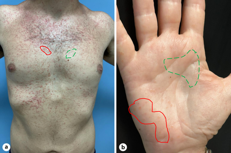
Introduction: Darier disease is a rare inherited disease with dominant skin manifestations including keratotic papules and plaques on sebaceous and flexural areas. Secondary infection of skin lesions is common, and Staphylococcus aureus commonly colonizes these lesions. The aim of the study was to characterize the bacterial microbiome of cutaneous Darier lesions compared to normal-looking skin and disease severity.

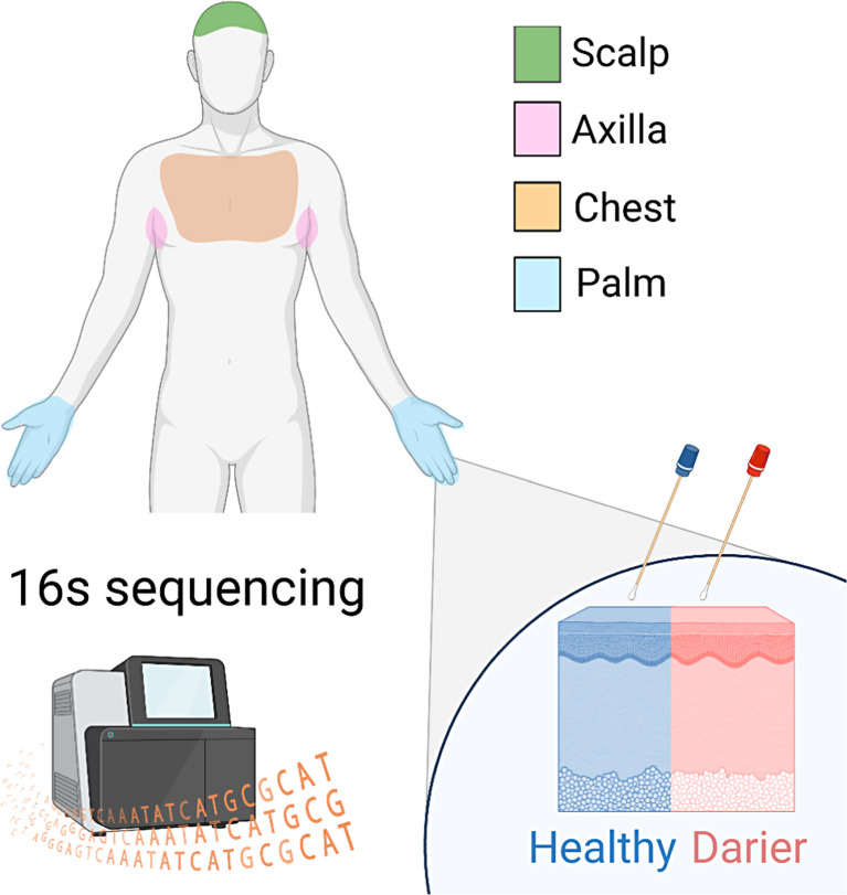
Methods: All patients with a history of Darier followed up at Emek Medical Center were invited to participate in the study. Patients that did not use antibiotics in the past month and signed informed consent had four skin sites sampled with swabs: scalp, chest, axilla, and palm. All samples were analyzed for bacterial microbiome using 16S rDNA sequencing.

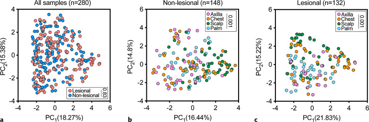
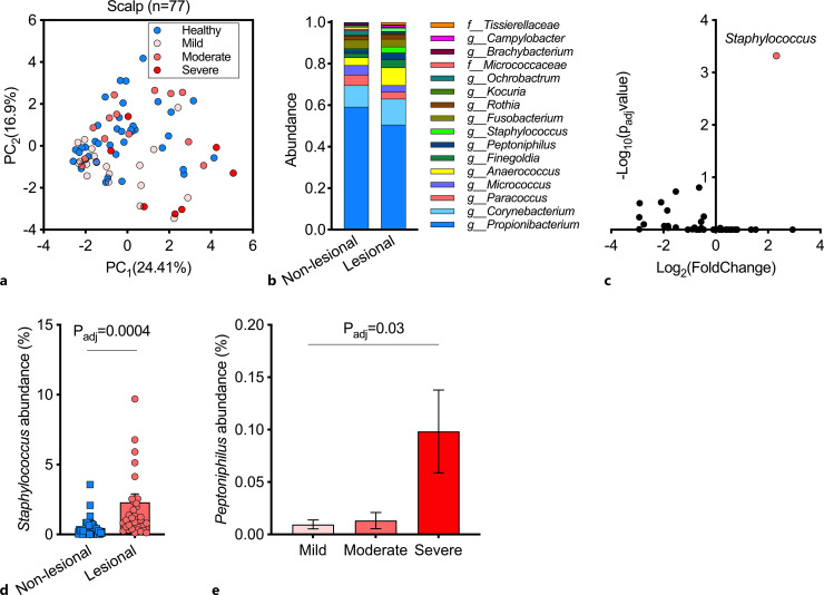
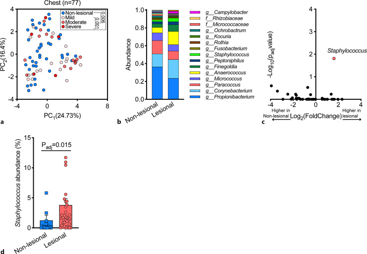
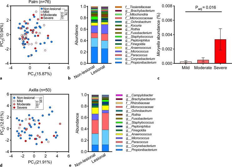
Results: Two hundred and eighty microbiome samples obtained from lesional and non-lesional skin of the scalp, chest, axilla, and palm of 42 Darier patients were included in the analysis. The most abundant bacterial genera across all skin sites were Propionibacterium, Corynebacterium, Paracoccus, Micrococcus, and Anaerococcus. Scalp and chest lesions featured a distinct microbiome configuration that was mainly driven by an overabundance of Staphylococci species. Patients with more severe disease exhibited microbiome alterations in the chest, axilla, and palm compared with patients with only mild disease, driven by Peptoniphilus and Moryella genera in scalp and palmar lesions, respectively.

Conclusion: Staphylococci were significantly associated with Darier lesions and drove Darier-associated dysbiosis. Severity of the disease was associated with two other bacterial genera. Whether these associations also hold a causative role and may serve as a therapeutic target remains to be determined and requires further investigation.

Keywords: Darier disease; Microbiome; Moryella; Peptoniphilus; Staphylococcus.


Conflict of interest statement

The authors state that there is no conflict of interest.

Figures

Fig. 1.
Fig. 1.
A representative scheme of the study design. We sampled the cutaneous microbiome from Darier patients using swabs in four body sites. For each Darier lesion, a control sample was collected from adjacent healthy-looking skin. All samples were sequenced by 16S rDNA sequencing.
Fig. 2.
Fig. 2.
Lesional versus non-lesional skin. An example of a classic study participant’s torso (a) and palm (b). The areas sampled as lesional skin are circled with a red straight curve and the areas sampled as non-lesional skin are circled by a green dotted curve.
Fig. 3.
Fig. 3.
Genus-level cutaneous microbiome composition of four body sites in patients with Darier. a A principal component analysis (PCA) of samples that were obtained from lesional and non-lesional skin. Inset: p value; permutational multivariate analysis of variance (PERMANOVA). b A PCA of microbiome composition in lesional skin, stratified by body site. Inset: p value; PERMNOVA. c A PCA of microbiome composition of non-lesional skin stratified by body site. Inset: p value; PERMANOVA. PC, principal component.
Fig. 4.
Fig. 4.
Cutaneous microbiome of the scalp in patients with Darier. a A principal component analysis (PCA) of samples that were obtained from lesional and non-lesional scalp skin. b Relative abundance of bacterial genera in samples from lesional and non-lesional scalp skin. c A differential abundance analysis of lesional and non-lesional scalp cutaneous microbiome. Two-sided Wilcoxon test, false discovery rate (FDR) correction for multiple comparisons (q < 0.05). d Distribution of staphylococcus abundance across lesional and non-lesional samples in scalp samples. Two-sided Wilcoxon test, false discovery rate (FDR) correction for multiple comparisons. e Relative abundance of Peptoniphilus as a function of disease severity (one-way analysis of variance comparing the mean abundance across all three categories, corrected by FDR for multiple comparisons of all genera). PC, principal component.
Fig. 5.
Fig. 5.
Cutaneous microbiome of the chest area in patients with Darier. a A PCA of 77 samples obtained from lesional and non-lesional skin of the chest. Inset: p value PERMANOVA. b Relative abundance of bacterial genera in samples from lesional and non-lesional chest skin. c A differential abundance analysis of lesional and non-lesional chest skin microbiome (two-sided Wilcoxon test, false discovery rate (FDR) correction for multiple comparisons (p < 0.05). d Distribution of staphylococcus abundance across lesional and non-lesional samples in chest samples. Two-sided Wilcoxon test, false discovery rate (FDR) correction for multiple comparisons. PC, principal component.
Fig. 6.
Fig. 6.
Cutaneous microbiome of the palm and axilla in patients with Darier. a A PCA of 76 samples obtained from lesional and non-lesional palmar skin. b Relative abundance of bacterial genera in samples from lesional and non-lesional palmar skin. c Relative abundance of Moryella in lesional skin across disease severity (one-way ANOVA). d A PCA of 50 samples obtained from lesional and non-lesional axillary skin. e Relative abundance of bacterial genera in samples from lesional and non-lesional axillary skin. PC, principal component.

References

    1. Carmona-Cruz S, Orozco-Covarrubias L, Sáez-de-Ocariz M. The human skin microbiome in selected cutaneous diseases. Front Cell Infect Microbiol. 2022;12:834135. - PMC - PubMed
    1. Byrd AL, Belkaid Y, Segre JA. The human skin microbiome. Nat Publ Gr. 2018;16(3):143–55. - PubMed
    1. Paller AS, Kong HH, Seed P, Naik S, Scharschmidt TC, Gallo RL, et al. . The microbiome in patients with atopic dermatitis. J Allergy Clin Immunol. 2019;143(1):26–35. - PMC - PubMed
    1. Kong HH, Oh J, Deming C, Conlan S, Grice EA, Beatson MA, et al. . Temporal shifts in the skin microbiome associated with disease flares and treatment in children with atopic dermatitis. Genome Res. 2012;22(5):850–9. - PMC - PubMed
    1. Brandwein M, Fuks G, Israel A, Sabbah F, Hodak E, Szitenberg A, et al. . Skin microbiome compositional changes in atopic dermatitis accompany dead sea climatotherapy. Photochem Photobiol. 2019;95(6):1446–53. - PubMed

MeSH terms