Clinical Trial
. 2024 Feb 20;5(2):101414.
doi: 10.1016/j.xcrm.2024.101414. Epub 2024 Feb 7.

Preclinical characterization and phase 1 results of ADG106 in patients with advanced solid tumors and non-Hodgkin's lymphoma

Affiliations
Clinical Trial

Preclinical characterization and phase 1 results of ADG106 in patients with advanced solid tumors and non-Hodgkin's lymphoma

Yuxiang Ma et al. Cell Rep Med. .

Abstract

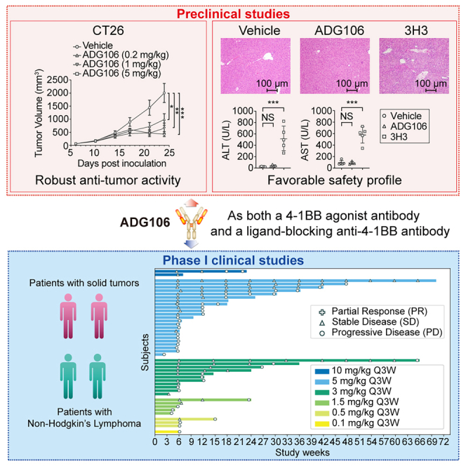
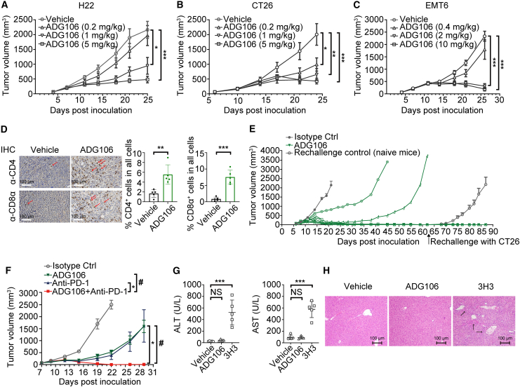
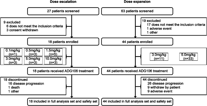
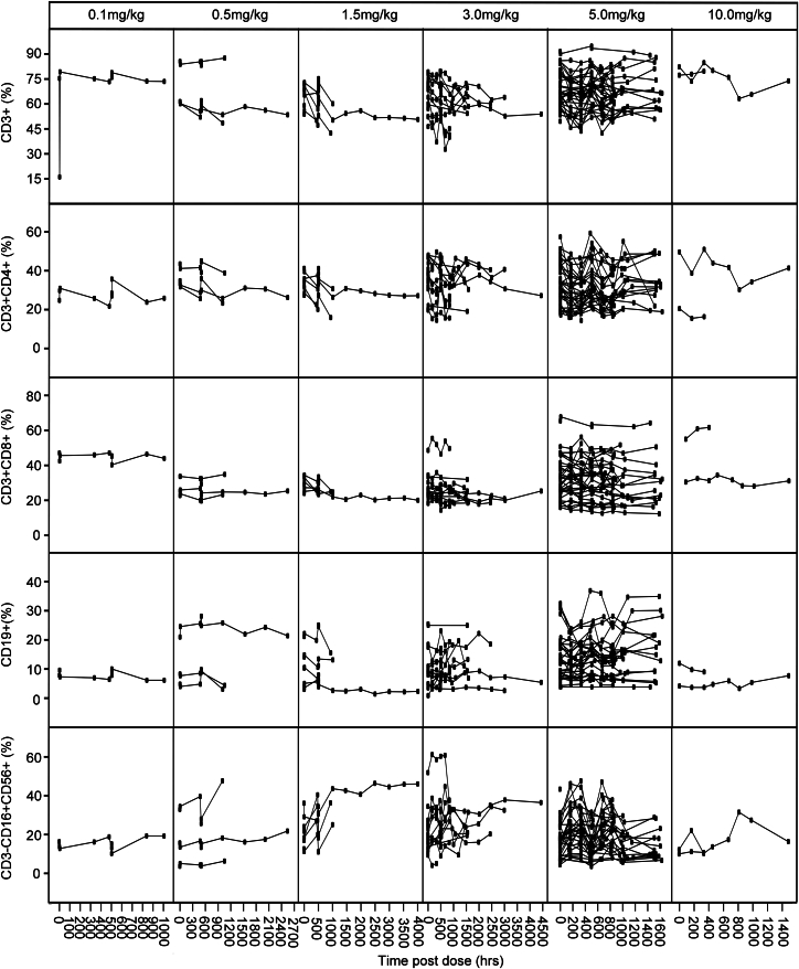
ADG106, a ligand-blocking agonistic antibody targeting CD137 (4-1BB), exhibits promising results in preclinical studies, demonstrating tumor suppression in various animal models and showing a balanced profile between safety and efficacy. This phase 1 study enrolls 62 patients with advanced malignancies, revealing favorable tolerability up to the 5.0 mg/kg dose level. Dose-limiting toxicity occurs in only one patient (6.3%) at 10.0 mg/kg, resulting in grade 4 neutropenia. The most frequent treatment-related adverse events include leukopenia (22.6%), neutropenia (22.6%), elevated alanine aminotransferase (22.6%), rash (21.0%), itching (17.7%), and elevated aspartate aminotransferase (17.7%). The overall disease control rates are 47.1% for advanced solid tumors and 54.5% for non-Hodgkin's lymphoma. Circulating biomarkers suggest target engagement by ADG106 and immune modulation of circulating T, B, and natural killer cells and cytokines interferon γ and interleukin-6, which may affect the probability of clinical efficacy. ADG106 has a manageable safety profile and preliminary anti-tumor efficacy in patients with advanced cancers (this study was registered at ClinicalTrials.gov: NCT03802955).

Keywords: ADG106; CD137/4-1BB; non-Hodgkin’s lymphoma; phase 1 trial; preclinical study; solid tumors.


Conflict of interest statement

Declaration of interests The authors declare no competing interests.

Figures

None
Graphical abstract
Figure 1
Figure 1
ADG106 exhibited its ability to bind activated CD4/CD8+ T cells, to activate NF-κB signaling, and to enhance T cell activation (A) ADG106 blocks interactions between CD137 and its ligand in a concentration-dependent manner. (B) Representative staining signals of ADG106 at different concentrations in CD4+ and CD8+ T cells from one donor are plotted. MFI, mean fluorescence intensity. Data are representative of five independent donors. (C) ADG106 stimulates CD137 receptor signaling upon FcγR-dependent crosslinking. Human CD137-expressing Jurkat/NF-κB-luciferase reporter cells were cocultured with CHO-K1 cells expressing human FcγRIIB and incubated with serially diluted human IgG4 isotype control antibody or ADG106 for 6 h. Activation of NF-κB signaling was detected by measuring the luciferase activity. (D and E) The CD137-expressing NF-κB-Luc Jurkat reporter cells were stimulated with the anti-CD137 antibodies in the presence (D) or absence (E) of cocultured CHO-K1 cells expressing human FcγRIIB. Luciferase activity was measured by bioluminescence assay. (F and G) ADG106 enhances T cell activation in the presence of suboptimal anti-CD3 stimulation. Human CD8+ T cells isolated from healthy donor were cultured on a 96-well plate precoated with anti-CD3 (1 mg/mL) and serially diluted ADG106 (starting from top 10 mg/mL at 10-fold dilution) for 96 h. Data are representative of two independent donors. T cell proliferation was measured by CellTiter-Glo and IFN-γ secretion by activated T cells in the supernatant was measured by ELISA. uns, unstimulated, in which no antibody was added including anti-CD3.
Figure 2
Figure 2
ADG106 inhibits tumor growth and is well tolerated in preclinical animal models (A–C) ADG106 induces dose-dependent anti-tumor efficacy in syngeneic murine H22 liver cancer model (A; n = 8), CT26 colon cancer model (B; n = 8), and EMT6 breast cancer model (C; n = 8). (D) Representative immunohistochemistry (IHC) staining images (200× magnification) of mouse CD4+ (top) and CD8α+ (bottom) T cells in H22 tumors after treatment with the vehicle control (left) or 5.0 mg/kg ADG106 antibody (right) (n = 5). CD4+ or CD8α+ T cells were stained in brown as indicated by arrows in the background of nuclear counterstain by hematoxylin. The percentages of CD4+ or CD8α+ T cells in all cells of the tumors treated with vehicle or ADG106 are plotted. ∗p < 0.05, ∗∗p < 0.01, and ∗∗∗p < 0.001. (E) BALB/c mice with established subcutaneous CT26 tumor were randomized into two treatment groups with 8 mice per group and treated with isotype control or ADG106 on day 6 at 5.0 mg/kg, intraperitoneally (i.p.) twice a week for 2 weeks. 6 of 8 mice in the ADG106-treated group exhibited complete tumor regression, and these mice were rechallenged again with CT26 tumor cells on day 62. Naive mice were also inoculated with CT26 tumor cells on the same day as rechallenge control. The tumor growth kinetics was monitored, with group mean ± SEM tumor volumes plotted for the isotype control and rechallenge control groups and with tumor volume for each individual mouse plotted for the ADG106 treatment group. (F) ADG106 was combined with a C57BL/6 mouse crossreactive anti-PD-1 antibody in the treatment of syngeneic Lewis lung cancer model. Mice with established subcutaneous tumors received i.p. dosing of ADG106 (5.0 mg/kg), anti-PD-1 (5.0 mg/kg), or their combination as indicated, twice a week for 2 weeks. Tumor volumes were monitored and plotted with group mean ± SEM (n = 8). ∗ or #p < 0.05. (G) Repeat-dose toxicity of ADG106 or 3H3 was conducted in normal BALB/c mice. Vehicle (n = 5), ADG106 (10.0 mg/kg; n = 4), or 3H3 (10.0 mg/kg; n = 5) was administered i.p., once weekly (QW) ±3 on days 0, 7, and 14. On day 28, animals were euthanized for postmortem examination and other analysis. Blood was collected from each animal for blood biochemistry (ALT, AST) analysis. The liver from each mouse was collected for histopathology analysis. ALT (left graph) and AST (right graph) levels of the BALB/c mice after treatment with vehicle, ADG106, and 3H3. (H) Representative histopathology images of liver sections from the mice treated with vehicle, ADG106, or 3H3. Immune cell infiltrations are indicated by arrows.
Figure 3
Figure 3
Study subjects included in the clinical study process
Figure 4
Figure 4
Waterfall plot of best percentage of change from baseline in the sum of diameters for target lesions and the swim plot indicating time to response in subjects accepting ADG106 treatment (A and B) Waterfall plot of best percentage of change from baseline in the sum of diameters for target lesions by in patients with solid tumors (A) and patients with non-Hodgkin’s lymphoma (B). Dashed lines indicate a 30% decrease and a 20% increase from baseline in the sum of longest diameters for target lesions. Patients with no measurable disease or no adequate baseline/post-baseline target lesion assessments were not included in this analysis. (C) Swim plot of duration of tumor response in patients with solid tumors and NHL. See also Table S1.
Figure 5
Figure 5
Changes in CD3+, CD3+CD4+, and CD3+CD8+ T cells, CD19+ B cells, and CD3CD16+CD56+ NK cells levels detected from day 1 of cycle 1 in individual patients by treatment group

References

    1. Shin D.S., Ribas A. The evolution of checkpoint blockade as a cancer therapy: what's here, what's next? Curr. Opin. Immunol. 2015;33:23–35. doi: 10.1016/j.coi.2015.01.006. - DOI - PubMed
    1. Moran A.E., Kovacsovics-Bankowski M., Weinberg A.D. The TNFRs OX40, 4-1BB, and CD40 as targets for cancer immunotherapy. Curr. Opin. Immunol. 2013;25:230–237. doi: 10.1016/j.coi.2013.01.004. - DOI - PMC - PubMed
    1. Antonia S.J., Larkin J., Ascierto P.A. Immuno-oncology combinations: a review of clinical experience and future prospects. Clin. Cancer Res. 2014;20:6258–6268. doi: 10.1158/1078-0432.Ccr-14-1457. - DOI - PubMed
    1. Segal N.H., He A.R., Doi T., Levy R., Bhatia S., Pishvaian M.J., Cesari R., Chen Y., Davis C.B., Huang B., et al. Phase I Study of Single-Agent Utomilumab (PF-05082566), a 4-1BB/CD137 Agonist, in Patients with Advanced Cancer. Clin. Cancer Res. 2018;24:1816–1823. doi: 10.1158/1078-0432.Ccr-17-1922. - DOI - PubMed
    1. Lynch D.H. The promise of 4-1BB (CD137)-mediated immunomodulation and the immunotherapy of cancer. Immunol. Rev. 2008;222:277–286. doi: 10.1111/j.1600-065X.2008.00621.x. - DOI - PubMed

Publication types

Substances

Associated data