

Polydopamine-assisted aptamer-carrying tetrahedral DNA microelectrode sensor for ultrasensitive electrochemical detection of exosomes
- PMID: 38331774
- PMCID: PMC10854160
- DOI: 10.1186/s12951-024-02318-6
Polydopamine-assisted aptamer-carrying tetrahedral DNA microelectrode sensor for ultrasensitive electrochemical detection of exosomes
Erratum in
-
Correction: Polydopamine-assisted aptamer-carrying tetrahedral DNA microelectrode sensor for ultrasensitive electrochemical detection of exosomes.J Nanobiotechnology. 2024 Feb 28;22(1):82. doi: 10.1186/s12951-024-02347-1. J Nanobiotechnology. 2024. PMID: 38419041 Free PMC article. No abstract available.
Abstract
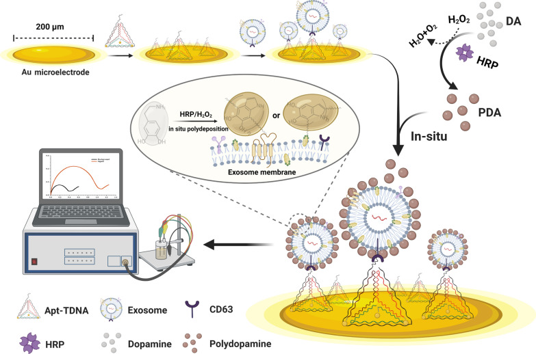
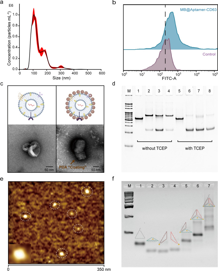
Background: Exosomes are nanoscale extracellular vesicles (30-160 nm) with endosome origin secreted by almost all types of cells, which are considered to be messengers of intercellular communication. Cancerous exosomes serve as a rich source of biomarkers for monitoring changes in cancer-related physiological status, because they carry a large number of biological macromolecules derived from parental tumors. The ultrasensitive quantification of trace amounts of cancerous exosomes is highly valuable for non-invasive early cancer diagnosis, yet it remains challenging. Herein, we developed an aptamer-carrying tetrahedral DNA (Apt-TDNA) microelectrode sensor, assisted by a polydopamine (PDA) coating with semiconducting properties, for the ultrasensitive electrochemical detection of cancer-derived exosomes.
Results: The stable rigid structure and orientation of Apt-TDNA ensured efficient capture of suspended exosomes. Without PDA coating signal amplification strategy, the sensor has a linear working range of 102-107 particles mL-1, with LOD of ~ 69 exosomes and ~ 42 exosomes for EIS and DPV, respectively. With PDA coating, the electrochemical signal of the microelectrode is further amplified, achieving single particle level sensitivity (~ 14 exosomes by EIS and ~ 6 exosomes by DPV).
Conclusions: The proposed PDA-assisted Apt-TDNA microelectrode sensor, which integrates efficient exosome capture, sensitive electrochemical signal feedback with PDA coating signal amplification, provides a new avenue for the development of simple and sensitive electrochemical sensing techniques in non-invasive cancer diagnosis and monitoring treatment response.
Keywords: Aptamer; Electrochemical sensing; Exosomes; Microelectrode; Polydopamine; Tetrahedra DNA.
© 2024. The Author(s).
Conflict of interest statement
The authors declare that they have no competing interests.
Figures


References
-
- Chia BS, Low YP, Wang Q, Li P, Gao ZQ. Advances in exosome quantification techniques. TrAC Trends Anal Chem. 2017;86:93–106. doi: 10.1016/j.trac.2016.10.012. - DOI
MeSH terms
Substances
Grants and funding
- 2022A1515011721/Natural Science Foundation of Guangdong Province
- 2022A1515010370/Natural Science Foundation of Guangdong Province
- 2019QN01Y725/Guangdong Provincial Pearl River Talents Program
- 202206010108/Science and Technology Program of Guangzhou
- 2020Z093/The Major Program of Ningbo Science and Technology Innovation 2025