. 2024 Feb 9;17(1):8.
doi: 10.1186/s13045-024-01525-w.

IL-15-secreting CAR natural killer cells directed toward the pan-cancer target CD70 eliminate both cancer cells and cancer-associated fibroblasts

Affiliations

IL-15-secreting CAR natural killer cells directed toward the pan-cancer target CD70 eliminate both cancer cells and cancer-associated fibroblasts

Astrid Van den Eynde et al. J Hematol Oncol. .

Abstract

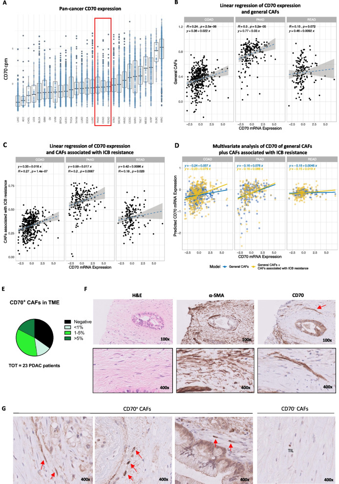
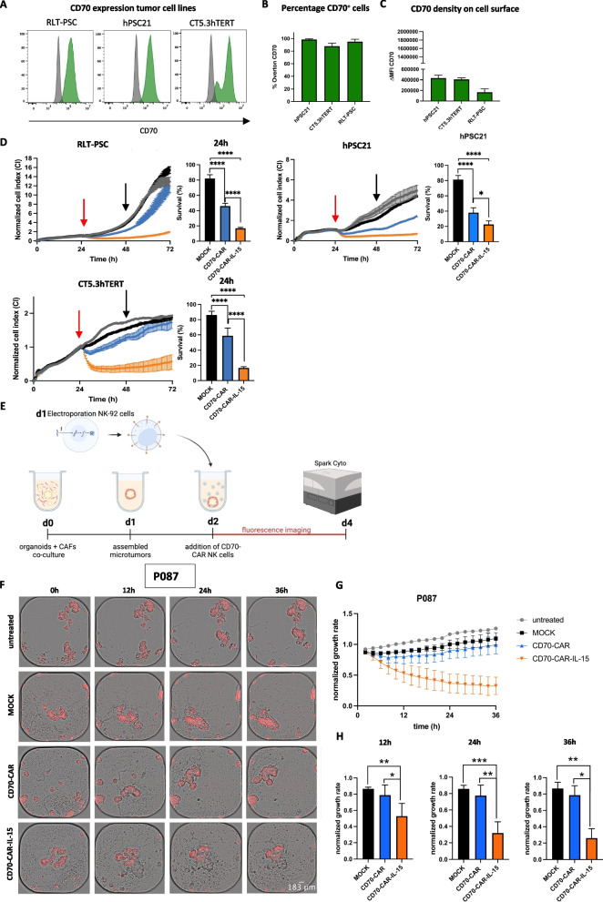
Background: It remains challenging to obtain positive outcomes with chimeric antigen receptor (CAR)-engineered cell therapies in solid malignancies, like colorectal cancer (CRC) and pancreatic ductal adenocarcinoma (PDAC). A major obstacle is the lack of targetable surface antigens that are not shared by healthy tissues. CD70 emerges as interesting target, due to its stringent expression pattern in healthy tissue and its apparent role in tumor progression in a considerable amount of malignancies. Moreover, CD70 is also expressed on cancer-associated fibroblasts (CAFs), another roadblock for treatment efficacy in CRC and PDAC. We explored the therapeutic potential of CD70 as target for CAR natural killer (NK) cell therapy in CRC, PDAC, focusing on tumor cells and CAFs, and lymphoma.

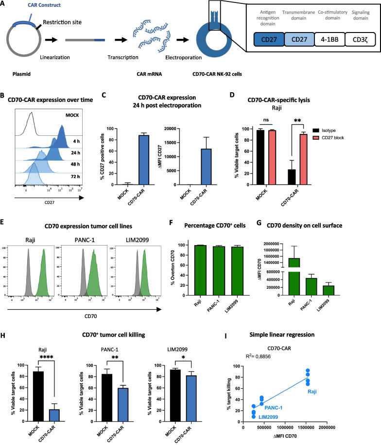
Methods: RNA-seq data and immunohistochemical analysis of patient samples were used to explore CD70 expression in CRC and PDAC patients. In addition, CD70-targeting CAR NK cells were developed to assess cytotoxic activity against CD70+ tumor cells and CAFs, and the effect of cytokine stimulation on their efficacy was evaluated. The in vitro functionality of CD70-CAR NK cells was investigated against a panel of tumor and CAF cell lines with varying CD70 expression. Lymphoma-bearing mice were used to validate in vivo potency of CD70-CAR NK cells. Lastly, to consider patient variability, CD70-CAR NK cells were tested on patient-derived organoids containing CAFs.

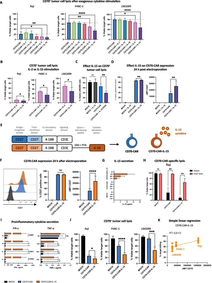
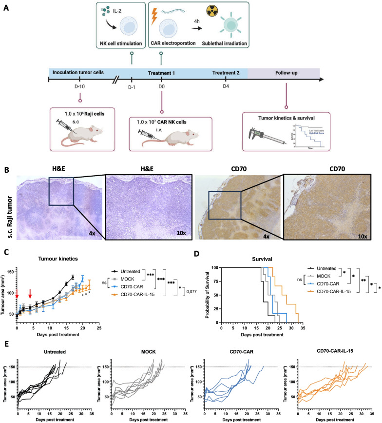
Results: In this study, we identified CD70 as a target for tumor cells and CAFs in CRC and PDAC patients. Functional evaluation of CD70-directed CAR NK cells indicated that IL-15 stimulation is essential to obtain effective elimination of CD70+ tumor cells and CAFs, and to improve tumor burden and survival of mice bearing CD70+ tumors. Mechanistically, IL-15 stimulation resulted in improved potency of CD70-CAR NK cells by upregulating CAR expression and increasing secretion of pro-inflammatory cytokines, in a mainly autocrine or intracellular manner.

Conclusions: We disclose CD70 as an attractive target both in hematological and solid tumors. IL-15 armored CAR NK cells act as potent effectors to eliminate these CD70+ cells. They can target both tumor cells and CAFs in patients with CRC and PDAC, and potentially other desmoplastic solid tumors.


Conflict of interest statement

The authors declare no competing interests.

Figures

Fig. 1
Fig. 1
CD70 expression in CRC and PDAC patients. A Pan-cancer CD70 expression in count per million reads (cpm) obtained from bulk RNA-seq of 28 different cancer types from the PCAWG Firehose cohort from the TCGA database. B, C Linear regression analysis and Spearman’s correlation analysis of CD70 mRNA expression and the general CAF score on CRC (i.e., COAD/READ) and PDAC (i.e., PAAD) databases, respectively. D Multivariate regression analysis with log ratio test to predict CD70 using the general CAF score (blue) and CAF score related to ICB therapy resistance on top of the general CAF score (yellow). E Distribution of percentage CD70+ CAFs of the total stromal fibroblasts found in the TME of 23 PDAC patients. F Representative images of H&E, α-SMA, and CD70 staining of a PDAC patient. G Representative images of CD70+ CAFs (indicated by red arrows) in the stromal compartment and an area with CD70 CAFs in the stromal compartment with a CD70+ TIL as reference. Total magnification is indicated in the lower right corner. Abbreviations: CRC, colorectal cancer; PDAC, pancreatic ductal adenocarcinoma; CAF, cancer-associated fibroblast; ICB, immune checkpoint blockade; TME, tumor microenvironment; H&E, hematoxylin and eosin; α-SMA, alpha-smooth muscle actin; and TIL, tumor-infiltrating lymphocyte
Fig. 2
Fig. 2
CD70-CAR NK cell development and validation. A Schematic representation of CD70-CAR NK cell generation and structural composition of the CD70-CAR construct. B CD70-CAR expression was detected by measuring CD27 expression on the cell surface using flow cytometry. Representative histograms of CD27 expression on NK-92 cells 4 h, 24 h, 48 h and 72 h after electroporation without (MOCK; white) or with CAR-encoding mRNA (CD70-CAR; blue). C Quantification of the amount CD27+ cells and the intensity of CD27 expression, depicted as mean fluorescence intensity minus isotype control (ΔMFI), 24 h post-electroporation (n = 6). D Percentage of viable Raji cells after a 4 h co-culture with CD70-CAR NK cells or MOCK control cells in the presence of 10 μg/mL anti-CD27 blocking antibody (CD27-block; red) or corresponding isotype control (Isotype; black; n = 4). E Representative flow cytometry histograms of CD70 expression on tumor cell lines (Raji, PANC-1, and LIM2099). F-G Quantification of percentage CD70+ cells and intensity of CD70 expression (ΔMFI) for Raji, PANC-1, and LIM2099 tumor cell lines, respectively. H Percentage of viable CD70+ tumor cells (Raji, PANC-1 and LIM2099) after a 4 h co-culture with CD70-CAR NK cells or MOCK control cells (n = 5). I Simple linear regression analysis of CD70-CAR NK cell target lysis and density of CD70 expression (ΔMFI) on target cells. Spearman’s correlation was used to analyze the correlation between the CD70 expression and target lysis. Linear mixed models were used to compare means of the lysis of the different tumor targets. ns = p > 0.05; *p < 0.05, **p < 0.01 and ****p < 0.0001
Fig. 3
Fig. 3
Effect of IL-15 cytokine stimulation on CD70-CAR NK cell functionality. A Percentage viable CD70+ target cells after a 4 h co-culture with unstimulated CD70-CAR NK cells or stimulated overnight with IL-2, IL-7, IL-12, IL-15, or IL-18 cytokines (n = 4). B Percentage viable CD70+ target cells after a 4 h co-culture with CD70-CAR NK cells stimulated overnight with IL-2 or IL-15 (n = 4). C Percentage viable LIM2099 cells after a 4 h co-culture with unstimulated MOCK or CD70-CAR NK cells or stimulated overnight with IL-15 (n = 5). D Quantification of CD27+ cells and intensity of CD27 expression (ΔMFI), 24 h after electroporation on CD70-CAR NK cells or MOCK cells cultured overnight with or without IL-15 (n = 5). E Structural design of CD70-CAR and CD70-CAR-IL-15 constructs. F Representative histograms and quantification of surface CD27 expression on MOCK NK cells (black), CD70-CAR NK cells (blue) and CD70-CAR-IL-15 NK cells (orange) 24 h after electroporation (n = 8). G Secreted IL-15 in the supernatant of MOCK, CD70-CAR NK cells, or CD70-CAR-IL-15 NK cells after electroporation (n = 3). H Percentage viable Raji cells after a 4 h co-culture with CD70-CAR NK cells, CD70-CAR-IL-15 NK cells or MOCK cells in the presence of 100 μg/mL anti-CD27 blocking antibody or isotype control (n = 4). I Secreted IFN-γ, and TNF-α in the supernatant of MOCK, CD70-CAR NK cells, or CD70-CAR-IL-15 NK cells after electroporation (n = 6). Error bars represent mean ± standard error of mean. J Percentage viable target cells after a 4 h co-culture with CD70-CAR NK cells, CD70-CAR-IL-15 NK cells or MOCK cells (n = 6). K Simple linear regression and Spearman Correlation analysis of CD70 ΔMFI and CD70-CAR-IL-15 target cell lysis. Linear mixed models with either Dunnett’s or Tukey’s correction for multiple comparison was applied to compare means. ns = p > 0.05; *p < 0.05, **p < 0.01, ***p < 0.001 and ****p < 0.0001
Fig. 4
Fig. 4
Tumor kinetics and survival of subcutaneous CD70+ Raji-bearing mice after treatment with CD70-CAR NK cells and CD70-CAR-IL-15 NK cells. A Schematic representation of (top) production of MOCK NK cells, CD70-CAR NK cells, and CD70-CAR-IL-15 NK cells prior to injection, and (bottom) schedule of generation and treatment of the CD70+ Raji xenograft mouse model. B Representative images of FFPE-slides of a Raji tumor stained for H&E and for CD70 positivity. Raji-bearing mice were treated twice with three days in between with 1.0 × 107 MOCK NK cells (n = 8), CD70-CAR NK cells (n = 6) or CD70-CAR-IL-15 NK cells (n = 6). Untreated mice were included as control (n = 8). C Tumor kinetics over time post-treatment; red arrows indicate treatment days. Error bars represent mean values ± standard error of mean. D Survival curve post-treatment. E Separated spider plots of the tumor kinetics over time per treatment group. Mixed model ANOVA was used to compare differences in tumor kinetics and the Log-Rank (Mantel-Cox) test was performed to analyze differences in survival. *p < 0.05, **p < 0.01 and ***p < 0.001
Fig. 5
Fig. 5
Cytotoxic activity of CD70-CAR NK cells and CD70-CAR-IL-15 NK cells against CD70+ CAFs. A-C CD70 expression on RLT-PSC, hPSC21, and CT5.3hTERT CAF cell lines. A Representative flow cytometry histograms. B-C Quantification of percentage CD70+ cells and intensity of CD70 expression (ΔMFI). D Representative graphs of longitudinal survival follow-up (i.e., Cell Index; CI) with the xCELLigence RTCA system and corresponding quantification after 24 h co-culture (indicated with black arrow; normalized to untreated) of RLT-PSC (n = 6), hPSC21 (n = 3), and CT5.3hTERT (n = 4) CAF cell lines with CD70-CAR NK cells, CD70-CAR-IL-15 NK cells, or MOCK NK cells. Monocultures of CAF cell lines treated with culture medium were used as control. To ensure proper adhesion, CAF cell lines were grown over 24 h after which treatment with (CAR) NK cells started (indicated by a red arrow) and the CI was followed up 48-h post-treatment. E Schematic representation of the experimental setup of PDAC patient-derived microtumors, containing tumor organoids with fluorescent red labelled RLT-PSC cells, in co-culture with (CAR) NK cells for two days. Monocultures of microtumors treated with medium were included as control. Follow up using live cell imaging was done using the Spark Cyto multimode reader. F–H Co-cultures of PDAC patient 087 microtumors (P87) with different treatment conditions: untreated, MOCK control NK cells, CD70-CAR NK cells, and CD70-CAR-IL-15 NK cells. F Representative brightfield images overlayed with the red fluorescent signal. G Growth rate, normalized against T0h, over time for the different treatment conditions. H Quantification of three different timepoints (12-h, 24-h, and 36-h post-treatment), comparing the treatment conditions normalized to the untreated control at that timepoint (n = 3). Images were cropped with ImageJ. Error bars represent mean values ± standard error of mean. Linear mixed models with Tukey’s correction for multiple comparison were applied to compare means of cell survival. * p < 0.05, **p < 0.01, ***p < 0.001 and ****p < 0.0001

References

    1. Flieswasser T, Camara-Clayette V, Danu A, Bosq J, Ribrag V, Zabrocki P, et al. Screening a broad range of solid and haematological tumour types for CD70 expression using a uniform IHC methodology as potential patient stratification method. Cancers (Basel). 2019;11(10):1611. doi: 10.3390/cancers11101611. - DOI - PMC - PubMed
    1. Kong F, Ye Q, Xiong Y. Comprehensive analysis of prognosis and immune function of CD70-CD27 signaling axis in pan-cancer. Funct Integr Genom. 2023;23:48. doi: 10.1007/s10142-023-00977-6. - DOI - PubMed
    1. Yang M, Tang X, Zhang Z, Gu L, Wei H, Zhao S, et al. Tandem CAR-T cells targeting CD70 and B7–H3 exhibit potent preclinical activity against multiple solid tumors. Theranostics. 2020;10(17):7622–7634. doi: 10.7150/thno.43991. - DOI - PMC - PubMed
    1. Borst J, Hendriks J, Xiao Y. CD27 and CD70 in T cell and B cell activation. Curr Opin Immunol. 2005;17(3):275–281. doi: 10.1016/j.coi.2005.04.004. - DOI - PubMed
    1. Jacobs J, Deschoolmeester V, Zwaenepoel K, Rolfo C, Silence K, Rottey S, et al. CD70: an emerging target in cancer immunotherapy. Pharmacol Ther. 2015;155:1–10. doi: 10.1016/j.pharmthera.2015.07.007. - DOI - PubMed

Publication types