

Cerebral MRI in a prospective cohort study on depression and atherosclerosis: the BiDirect sample, processing pipelines, and analysis tools
- PMID: 38332362
- PMCID: PMC10853141
- DOI: 10.1186/s41747-023-00415-z
Cerebral MRI in a prospective cohort study on depression and atherosclerosis: the BiDirect sample, processing pipelines, and analysis tools
Abstract
Background: The use of cerebral magnetic resonance imaging (MRI) in observational studies has increased exponentially in recent years, making it critical to provide details about the study sample, image processing, and extracted imaging markers to validate and replicate study results. This article reviews the cerebral MRI dataset from the now-completed BiDirect cohort study, as an update and extension of the feasibility report published after the first two examination time points.
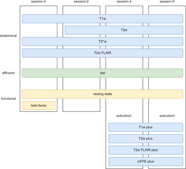
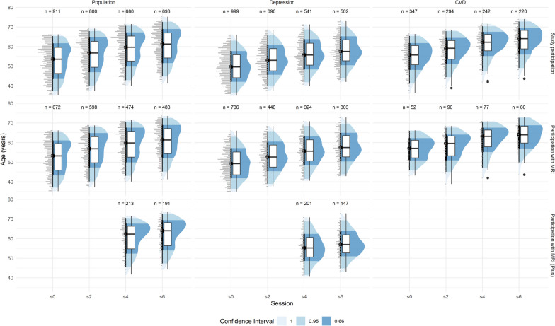
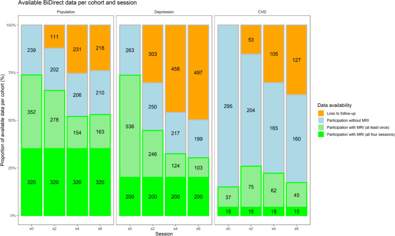
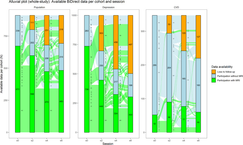
Methods: We report the sample and flow of participants spanning four study sessions and twelve years. In addition, we provide details on the acquisition protocol; the processing pipelines, including standardization and quality control methods; and the analytical tools used and markers available.
Results: All data were collected from 2010 to 2021 at a single site in Münster, Germany, starting with a population of 2,257 participants at baseline in 3 different cohorts: a population-based cohort (n = 911 at baseline, 672 with MRI data), patients diagnosed with depression (n = 999, 736 with MRI data), and patients with manifest cardiovascular disease (n = 347, 52 with MRI data). During the study period, a total of 4,315 MRI sessions were performed, and over 535 participants underwent MRI at all 4 time points.
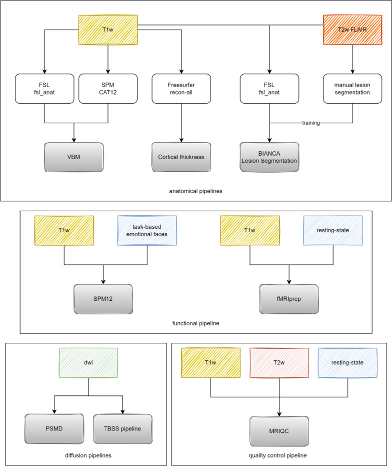
Conclusions: Images were converted to Brain Imaging Data Structure (a standard for organizing and describing neuroimaging data) and analyzed using common tools, such as CAT12, FSL, Freesurfer, and BIANCA to extract imaging biomarkers. The BiDirect study comprises a thoroughly phenotyped study population with structural and functional MRI data.
Relevance statement: The BiDirect Study includes a population-based sample and two patient-based samples whose MRI data can help answer numerous neuropsychiatric and cardiovascular research questions.
Key points: • The BiDirect study included characterized patient- and population-based cohorts with MRI data. • Data were standardized to Brain Imaging Data Structure and processed with commonly available software. • MRI data and markers are available upon request.
Keywords: Longitudinal studies; Magnetic resonance imaging; Medical image processing; Population health; Standardization.
© 2024. The Author(s).
Conflict of interest statement
The authors of this manuscript declare no relationships with any companies, whose products or services may be related to the subject matter of the article.
Figures
Similar articles
-
MR imaging of the brain in large cohort studies: feasibility report of the population- and patient-based BiDirect study.Eur Radiol. 2017 Jan;27(1):231-238. doi: 10.1007/s00330-016-4303-9. Epub 2016 Apr 8. Eur Radiol. 2017. PMID: 27059857
-
Establishing the bidirectional relationship between depression and subclinical arteriosclerosis--rationale, design, and characteristics of the BiDirect Study.BMC Psychiatry. 2014 Jun 13;14:174. doi: 10.1186/1471-244X-14-174. BMC Psychiatry. 2014. PMID: 24924233 Free PMC article.
-
[New cohorts. The BiDirect study].Bundesgesundheitsblatt Gesundheitsforschung Gesundheitsschutz. 2012 Jun;55(6-7):822-3. doi: 10.1007/s00103-012-1491-6. Bundesgesundheitsblatt Gesundheitsforschung Gesundheitsschutz. 2012. PMID: 22736162 German.
-
Structural magnetic resonance imaging for the early diagnosis of dementia due to Alzheimer's disease in people with mild cognitive impairment.Cochrane Database Syst Rev. 2020 Mar 2;3(3):CD009628. doi: 10.1002/14651858.CD009628.pub2. Cochrane Database Syst Rev. 2020. PMID: 32119112 Free PMC article.
-
Contrast agent-free state-of-the-art magnetic resonance imaging on cerebral small vessel disease - Part 2: Diffusion tensor imaging and functional magnetic resonance imaging.NMR Biomed. 2022 Aug;35(8):e4743. doi: 10.1002/nbm.4743. Epub 2022 May 6. NMR Biomed. 2022. PMID: 35429070 Review.
References
Publication types
MeSH terms
Grants and funding
LinkOut - more resources
Full Text Sources
Medical