. 2024 Jan 25:15:1327948.
doi: 10.3389/fphys.2024.1327948. eCollection 2024.

AI algorithm for personalized resource allocation and treatment of hemorrhage casualties

Affiliations

AI algorithm for personalized resource allocation and treatment of hemorrhage casualties

Xin Jin et al. Front Physiol. .

Abstract

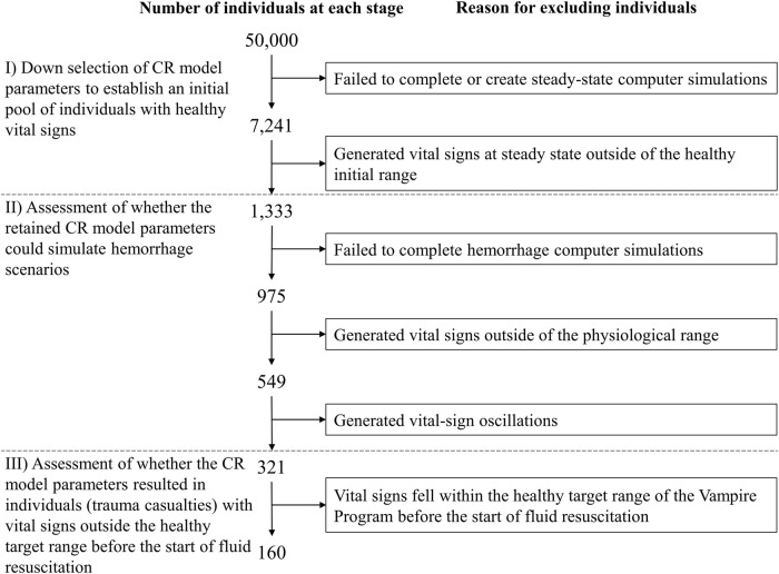
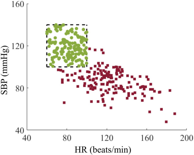
A deep neural network-based artificial intelligence (AI) model was assessed for its utility in predicting vital signs of hemorrhage patients and optimizing the management of fluid resuscitation in mass casualties. With the use of a cardio-respiratory computational model to generate synthetic data of hemorrhage casualties, an application was created where a limited data stream (the initial 10 min of vital-sign monitoring) could be used to predict the outcomes of different fluid resuscitation allocations 60 min into the future. The predicted outcomes were then used to select the optimal resuscitation allocation for various simulated mass-casualty scenarios. This allowed the assessment of the potential benefits of using an allocation method based on personalized predictions of future vital signs versus a static population-based method that only uses currently available vital-sign information. The theoretical benefits of this approach included up to 46% additional casualties restored to healthy vital signs and a 119% increase in fluid-utilization efficiency. Although the study is not immune from limitations associated with synthetic data under specific assumptions, the work demonstrated the potential for incorporating neural network-based AI technologies in hemorrhage detection and treatment. The simulated injury and treatment scenarios used delineated possible benefits and opportunities available for using AI in pre-hospital trauma care. The greatest benefit of this technology lies in its ability to provide personalized interventions that optimize clinical outcomes under resource-limited conditions, such as in civilian or military mass-casualty events, involving moderate and severe hemorrhage.

Keywords: artificial intelligence; fluid resuscitation; hemorrhage; resource utilization; trauma.


Conflict of interest statement

The authors declare that the research was conducted in the absence of any commercial or financial relationships that could be construed as a potential conflict of interest. The author(s) declared that they were an editorial board member of Frontiers, at the time of submission. This had no impact on the peer review process and the final decision.

Figures

FIGURE 1
FIGURE 1
Outline of the methodology used to develop and assess the artificial intelligence (AI) algorithm for personalized resource allocation of hemorrhage casualties. I) Synthetic-data generation: Use the cardio-respiratory (CR) model to perform simulations and generate synthetic trauma casualties with associated vital-sign trajectories, for a given hemorrhage-inducing trauma condition and each of four fluid treatment options. II) AI-model development: Using the CR-generated synthetic data, perform a 5-fold nested cross-validation to develop AI models that use 10 min of pre-fluid-treatment vital signs to predict vital signs 60 min into the future after fluid treatment. III) AI and Vampire assessment: Use the CR-generated vital-sign data to compare the outcomes in terms of the number of restored casualties to a “safe” physiological state and the amount of fluid utilization for the optimal fluid treatments allocated by the AI model and the Vampire Program as well as the CR-based optimal fluid treatment.
FIGURE 2
FIGURE 2
Events and time intervals used to create different scenarios representing an initial hemorrhage-inducing trauma, tourniquet application, and subsequent fluid resuscitation treatment. The injury at t0 is followed by a period of uncontrolled bleeding for a minimum of 5 min, after which a tourniquet is applied within a 10-min interval, i.e., from 5 to 15 min after the injury. The tourniquet application at t1 stops the bleeding, the fluid transfusion at t2 is initiated at a time interval 10–15 min after t1, and the transfusion continues for another 60 min until t3. Different scenarios sample different time intervals between t0 and t1 to apply a tourniquet and between t1 and t2 to start the transfusion, with blood transfusion starting between 15 and 30 min after the traumatic event. The maximum transfusion time is fixed at 60 min.
FIGURE 3
FIGURE 3
The range of bleeding times and blood-volume losses used to create different hemorrhage scenarios. The pentagon-shaded area defines the range of bleeding parameters used to create Class II and III hemorrhage cases (Schwartz and Holcomb, 2017) for this study, compatible with bleeding times of 5–15 min and blood-volume losses of 0.75–2.00 L. The blue dash-dotted edge of the pentagon represents the 0.22 L/min maximum rate of hemorrhage for these scenarios.
FIGURE 4
FIGURE 4
Transfusion rates of four fluid treatment options. 1) 30 min 0 unit + 30 min 0 unit; 2) 30 min 0 unit + 30 min 1 unit; 3) 30 min 1 unit + 30 min 0 unit; or 4) 30 min 1 unit + 30 min 1 unit. The fluid treatment starts at t2 and continues for 60 min until t3.
FIGURE 5
FIGURE 5
Procedure for selection of cardio-respiratory (CR) model parameter sets representing individuals used to generate vital-sign trajectories associated with the simulated hemorrhage and treatment scenarios. The selection procedure includes three different stages (I–III) in order to generate a broad range of individuals with vital signs in the healthy target range before hemorrhage and outside of this range after hemorrhage onset, to successfully simulate different degrees of moderate to severe hemorrhage.
FIGURE 6
FIGURE 6
Structure of the recurrent neural network AI model. The model’s inputs are the fluid infusion rate [uf(t)], heart rate [HR(t)], and systolic blood pressure [SBP(t)] at time t, and the outputs consist of the predicted heart rate [ HR^ (t+1)] and the predicted systolic blood pressure [ SBP^ (t+1)] for the subsequent minute. The model architecture includes two feedforward layers and a gated recurrent unit (GRU) layer, each with 512 nodes.
FIGURE 7
FIGURE 7
Distribution of heart rate (HR) and systolic blood pressure (SBP) for the cohort of 160 trauma casualties before (green circles) and after (red squares) hemorrhage. The range within the black dashed lines represents the healthy initial range.
FIGURE 8
FIGURE 8
Comparison of Vampire- and AI-based allocation methods for the single casualty in Analysis 1. (A) Heart rate (HR) and (B) systolic blood pressure (SBP) over time, where t1 denotes the time for tourniquet application and t2 represents the time for initiation of fluid resuscitation, where the horizontal black solid lines represent the boundaries of the healthy target range. The red solid lines represent vital signs during the hemorrhage phase, the yellow dashed lines denote vital signs with no fluid transfusion, and the green dash-dotted lines represent vital signs after receiving 1 unit of fluids at t2 infused for 30 min during the treatment phase.
FIGURE 9
FIGURE 9
Comparison of fluid allocations based on the cardio-respiratory (CR) model, AI predictions, and the Vampire Program for different numbers of available fluid units. (A) Number of casualties restored to the healthy target range. (B) Excessive use of fluid units (number of fluid units used more than required based on the gold-standard CR results). The shaded areas represent two standard errors of the mean.
FIGURE 10
FIGURE 10
Classification results of the linear support vector machine algorithm for vital-sign trajectories at the end of fluid resuscitation at time t3 for the scenarios 1) when tourniquet application at t1 controlled bleeding and 2) when tourniquet application at t1 did not control all bleeding because there was additional non-compressible bleeding. The blue circles and red squares represent the prediction errors between the CR and AI models (CR minus AI) for heart rate (HR) and systolic blood pressure (SBP) at t3 for the two scenarios. The blue and red shaded areas represent the classified areas corresponding to controlled bleeding and non-compressible bleeding, respectively. The red dashed line between these two areas denotes the decision boundary that separates the two scenarios.

Similar articles

Cited by

References

    1. Alsalti M., Tivay A., Jin X., Kramer G. C., Hahn J.-O. (2022). Design and in silico evaluation of a closed-loop hemorrhage resuscitation algorithm with blood pressure as controlled variable. J. Dyn. Syst. Meas. Control. 144, 021001. 10.1115/1.4052312 - DOI
    1. Avital G., Snider E. J., Berard D., Vega S. J., Hernandez Torres S. I., Convertino V. A., et al. (2022). Closed-loop controlled fluid administration systems: a comprehensive scoping review. J. Pers. Med. 12, 1168. 10.3390/jpm12071168 - DOI - PMC - PubMed
    1. Bray A., Webb J. B., Enquobahrie A., Vicory J., Heneghan J., Hubal R., et al. (2019). Pulse physiology engine: an open-source software platform for computational modeling of human medical simulation. SN Comp. Clin. Med. 1, 362–377. 10.1007/s42399-019-00053-w - DOI
    1. Burges C. J. (1998). A tutorial on support vector machines for pattern recognition. Data Min. Knowl. Discov. 2, 121–167. 10.1023/A:1009715923555 - DOI
    1. Chang R., Cardenas J. C., Wade C. E., Holcomb J. B. (2016). Advances in the understanding of trauma-induced coagulopathy. Blood 128, 1043–1049. 10.1182/blood-2016-01-636423 - DOI - PMC - PubMed

LinkOut - more resources