

Endodontic Treatment of a Maxillary First Molar With Two Separate Palatal Roots: A Case Report
- PMID: 38333498
- PMCID: PMC10850443
- DOI: 10.7759/cureus.51907
Endodontic Treatment of a Maxillary First Molar With Two Separate Palatal Roots: A Case Report
Abstract
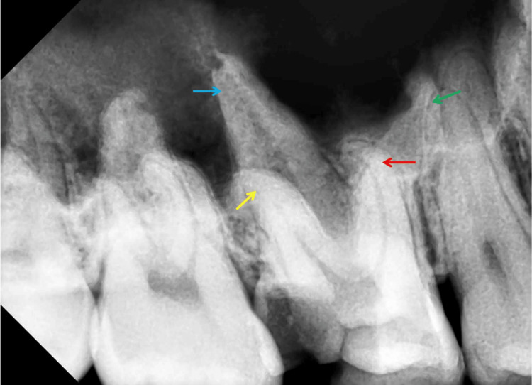
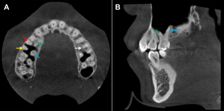
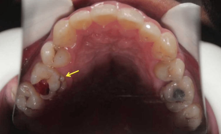
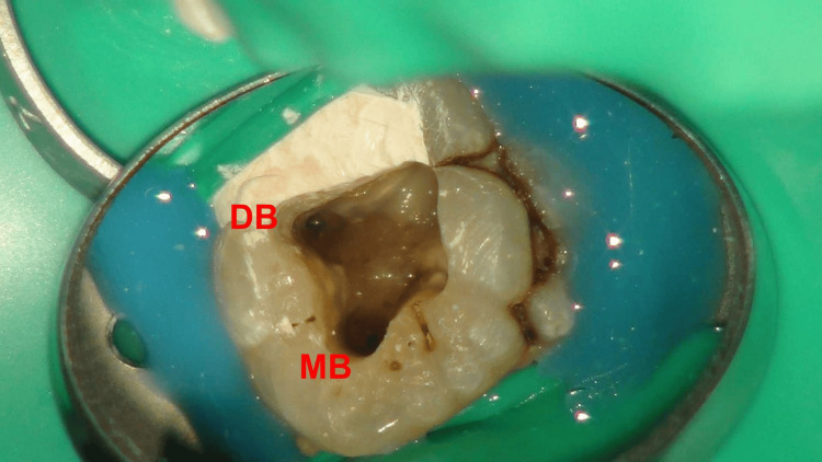
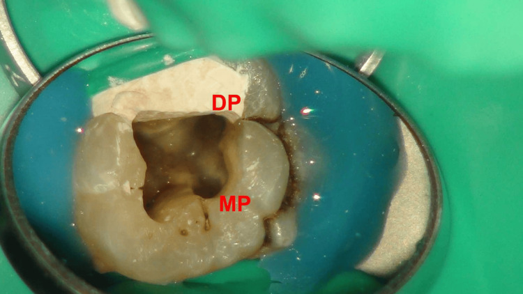
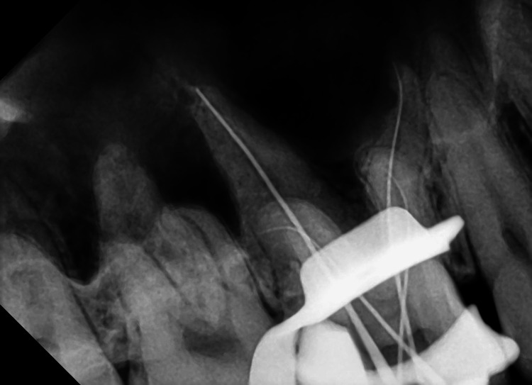
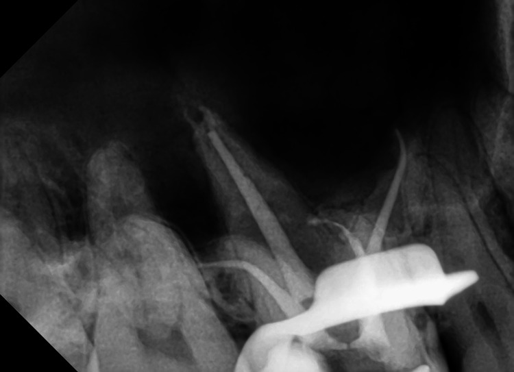
Maxillary first molars exhibit considerable anatomical variation, with a single palatal root being the most common occurrence, while two palatal roots are notably less frequent. This case report details the endodontic treatment of a maxillary first molar with two separate palatal roots. It highlights the critical importance of recognizing these anatomical variations and their unique challenges during endodontic procedures. This report emphasizes the essential role of advanced diagnostic methods, such as cone-beam computed tomography, and the use of microscopic techniques in identifying and treating such cases.
Keywords: cone- beam computed tomography; dental operating microscope; extra palatal root; maxillary first molar; root canal treatment.
Copyright © 2024, Liu et al.
Conflict of interest statement
The authors have declared that no competing interests exist.
Figures
Similar articles
-
Cone Beam Computed Tomography-aided Endodontic Management of Second Maxillary Molar with Two Separate Palatal Roots: A Case Report.Cureus. 2020 Mar 20;12(3):e7347. doi: 10.7759/cureus.7347. Cureus. 2020. PMID: 32226696 Free PMC article.
-
Endodontic treatment of various palatal roots in maxillary molars: Case series and clinical experience.J Am Dent Assoc. 2021 Dec;152(12):1044-1052. doi: 10.1016/j.adaj.2021.05.001. Epub 2021 Jul 24. J Am Dent Assoc. 2021. PMID: 34311979
-
Endodontic treatment of the maxillary first molar with palatal canal variations: A case report and review of literature.World J Clin Cases. 2022 Nov 16;10(32):12036-12044. doi: 10.12998/wjcc.v10.i32.12036. World J Clin Cases. 2022. PMID: 36405283 Free PMC article.
-
Variations of Palatal Canal Morphology in Maxillary Molars: A Case Series and Literature Review.J Endod. 2017 Nov;43(11):1888-1896. doi: 10.1016/j.joen.2017.04.006. Epub 2017 Jun 30. J Endod. 2017. PMID: 28673493 Review.
-
Evaluation of root canal morphology of human primary molars by using CBCT and comprehensive review of the literature.Acta Odontol Scand. 2016;74(4):250-8. doi: 10.3109/00016357.2015.1104721. Epub 2015 Nov 2. Acta Odontol Scand. 2016. PMID: 26523502 Review.
Cited by
-
Endodontic Treatment of a Maxillary Second Molar With Five Canals: A Case Report and a Literature Review.Cureus. 2024 Apr 27;16(4):e59179. doi: 10.7759/cureus.59179. eCollection 2024 Apr. Cureus. 2024. PMID: 38807838 Free PMC article.
-
Locating and Treating Three Calcified Canals in a Mandibular First Molar With Radix Entomolaris and Five Canals: A Case Report.Cureus. 2024 Jan 25;16(1):e52931. doi: 10.7759/cureus.52931. eCollection 2024 Jan. Cureus. 2024. PMID: 38406086 Free PMC article.
References
-
- Eradication of endodontic infection by instrumentation and irrigation solutions. Haapasalo M, Endal U, Zandi H, Coil JM. Endod topics. 2005;10:77–102.
-
- Missed anatomy: frequency and clinical impact. Cantatore G, Berutti E, Castellucci A. Endod Topics. 2006;15:3–31.
-
- Antimicrobial effects of agitational irrigation on single- and multispecies biofilms in dentin canals. Al-Zuhair H, Su Z, Liu H, et al. Odontology. 2023;111:49–56. - PubMed
Publication types
LinkOut - more resources
Full Text Sources