

18F-FDG-PET/CT for Localizing the Epileptogenic Focus in Patients with Different Types of Focal Cortical Dysplasia
- PMID: 38333612
- PMCID: PMC10849898
- DOI: 10.2147/NDT.S442459
18F-FDG-PET/CT for Localizing the Epileptogenic Focus in Patients with Different Types of Focal Cortical Dysplasia
Abstract
Purpose: To determine the diagnostic and localization value of 18F-fluorodeoxyglucose-positron emission tomography (PET)/computed tomography (CT) in patients with focal cortical dysplasia (FCD) who underwent epilepsy surgery.
Methods: One hundred and eight patients with pathologically proven FCD who underwent surgery for refractory epilepsy were retrospectively analyzed. All patients underwent magnetic resonance imaging (MRI), 18F-FDG-PET/CT, and video electroencephalography. An MRI diagnosis of FCD was defined as MRI+. A PET/CT diagnosis of FCD was defined as PET/CT+.
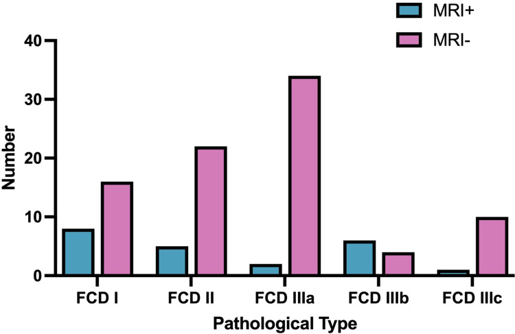
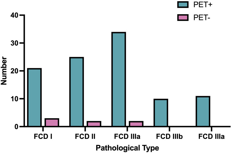
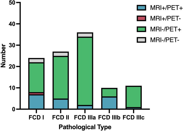
Results: MRI and PET/CT detected FCD in 20.37% and 93.52% of patients, respectively. The difference was significant. Twenty-one patients were MRI+/PET+, 80 were MRI-/PET+, six were MRI-/PET-, and one was MRI+/PET-. The MRI positivity rate was lowest in patients with FCD type IIIa (5.6%, P < 0.05). Prevalence of MRI-/PET+ was highest in patients with FCD type IIIa (88.89%, P < 0.05).
Conclusion: PET/CT is superior to MRI in detecting FCD. FCD type IIIa was more likely than other types to show MRI-/PET+. This suggests that PET/CT has particular diagnostic value for FCD type IIIa patients with negative MRI findings.
Keywords: 18F-FDG-PET/CT; epilepsy; focal cortical dysplasia; pathological types.
© 2024 Wang et al.
Conflict of interest statement
The authors have no competing interests.
Figures
Similar articles
-
[18F]SynVesT-1 and [18F]FDG quantitative PET imaging in the presurgical evaluation of MRI-negative children with focal cortical dysplasia type II.Eur J Nucl Med Mol Imaging. 2024 May;51(6):1651-1661. doi: 10.1007/s00259-024-06593-1. Epub 2024 Jan 6. Eur J Nucl Med Mol Imaging. 2024. PMID: 38182838
-
Cortical abnormalities of synaptic vesicle protein 2A in focal cortical dysplasia type II identified in vivo with 18F-SynVesT-1 positron emission tomography imaging.Eur J Nucl Med Mol Imaging. 2022 Aug;49(10):3482-3491. doi: 10.1007/s00259-021-05665-w. Epub 2022 Jan 3. Eur J Nucl Med Mol Imaging. 2022. PMID: 34978594 Free PMC article.
-
Compatibility of MRI and FDG-PET findings with histopathological results in patients with focal cortical dysplasia.Seizure. 2017 Feb;45:80-86. doi: 10.1016/j.seizure.2016.11.024. Epub 2016 Dec 6. Seizure. 2017. PMID: 27960132
-
Focal cortical dysplasia and epilepsy surgery.J Epilepsy Res. 2013 Dec 30;3(2):43-7. doi: 10.14581/jer.13009. eCollection 2013 Dec. J Epilepsy Res. 2013. PMID: 24649472 Free PMC article. Review.
-
Diagnostic methods and treatment options for focal cortical dysplasia.Epilepsia. 2015 Nov;56(11):1669-86. doi: 10.1111/epi.13200. Epub 2015 Oct 5. Epilepsia. 2015. PMID: 26434565 Review.
Cited by
-
A comparative analysis of imaging-based algorithms for detecting focal cortical dysplasia type II in children.Sci Rep. 2025 Aug 15;15(1):29946. doi: 10.1038/s41598-025-16015-3. Sci Rep. 2025. PMID: 40817382 Free PMC article.
References
LinkOut - more resources
Full Text Sources
