. 2024 Jun 3;26(6):1109-1123.
doi: 10.1093/neuonc/noae015.

Unraveling the complexity of the senescence-associated secretory phenotype in adamantinomatous craniopharyngioma using multimodal machine learning analysis

Affiliations

Unraveling the complexity of the senescence-associated secretory phenotype in adamantinomatous craniopharyngioma using multimodal machine learning analysis

Eric W Prince et al. Neuro Oncol. .

Abstract

Background: Cellular senescence can have positive and negative effects on the body, including aiding in damage repair and facilitating tumor growth. Adamantinomatous craniopharyngioma (ACP), the most common pediatric sellar/suprasellar brain tumor, poses significant treatment challenges. Recent studies suggest that senescent cells in ACP tumors may contribute to tumor growth and invasion by releasing a senesecence-associated secretory phenotype. However, a detailed analysis of these characteristics has yet to be completed.

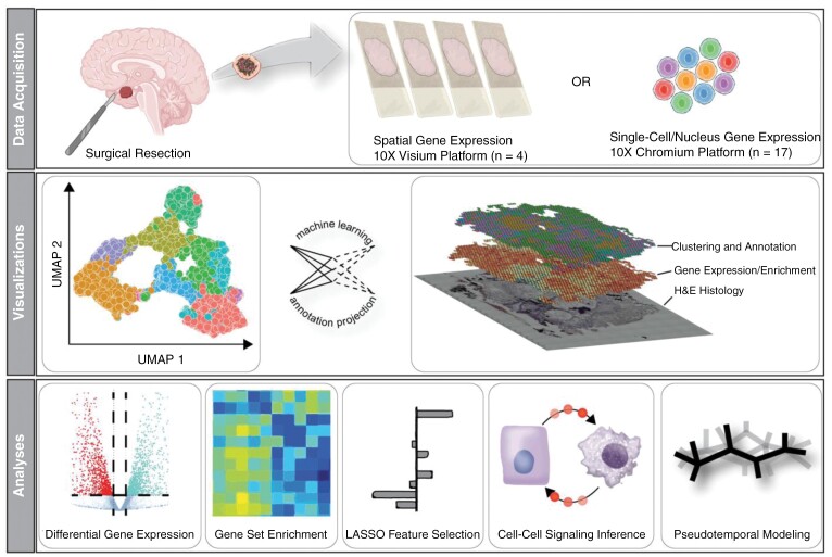
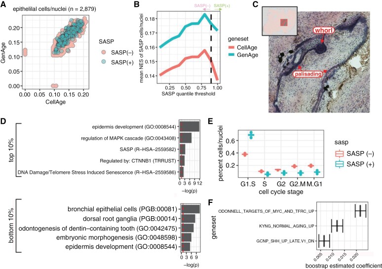
Methods: We analyzed primary tissue samples from ACP patients using single-cell, single-nuclei, and spatial RNA sequencing. We performed various analyses, including gene expression clustering, inferred senescence cells from gene expression, and conducted cytokine signaling inference. We utilized LASSO to select essential gene expression pathways associated with senescence. Finally, we validated our findings through immunostaining.

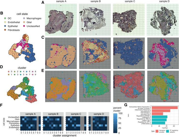
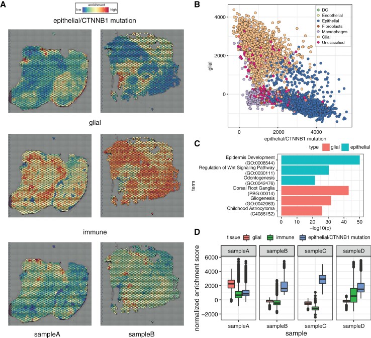
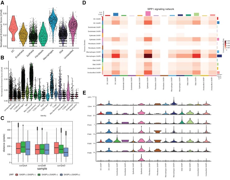
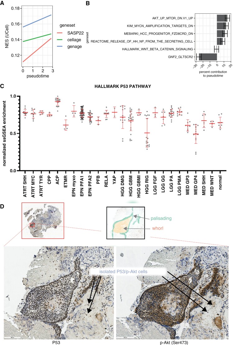
Results: We observed significant diversity in gene expression and tissue structure. Key factors such as NFKB, RELA, and SP1 are essential in regulating gene expression, while senescence markers are present throughout the tissue. SPP1 is the most significant cytokine signaling network among ACP cells, while the Wnt signaling pathway predominantly occurs between epithelial and glial cells. Our research has identified links between senescence-associated features and pathways, such as PI3K/Akt/mTOR, MYC, FZD, and Hedgehog, with increased P53 expression associated with senescence in these cells.

Conclusions: A complex interplay between cellular senescence, cytokine signaling, and gene expression pathways underlies ACP development. Further research is crucial to understand how these elements interact to create novel therapeutic approaches for patients with ACP.

Keywords: adamantinomatous craniopharyngioma (ACP); machine learning; next generation sequencing; pediatric neuro-oncology; senescence-associated secretory phenotype (SASP).


Conflict of interest statement

None declared.

Figures

Graphical Abstract
Graphical Abstract
Figure 1.
Figure 1.
Spatial transcriptional heterogeneity in adamantinomatous craniopharyngioma tissue. (a) Brightfield microscopy images for the four spatial gene expression samples acquired. (b, c). UMAP representation of spatial gene expression data annotated for inferred cell state with annotations overlayed on microscopy images. (d, e) UMAP representation of spatial gene expression data annotated for cluster ID with annotations overlayed on microscopy images. (f) Heatmap is stratifying the percentages of cluster cell state composition per patient. Note that all percentages are per column; a cluster typically corresponds to one cell type. (g) Enriched biological processes and transcription factor (TF) regulatory networks for spatially variable genes in ACP tissue.
Figure 2.
Figure 2.
Gene set enrichment identifies epithelial (eg, tumor), glial, and immune spatial gene expression compartments within ACP tissue samples. (a) Example images of single-sample gene set enrichment analysis for Apps epithelial/CTNNB1 mutation, glial, and immune gene sets, respectively; red spots indicate high enrichment, and blue spots indicate low enrichment. (b) Scatterplot of enrichment scores for epithelial/CTNNB1 mutation and glial modules; spots are colored by cell state as shown in Figure 1b. (c) Enriched terms (ontologies, pathways, processes, diseases, cell types) for gene markers identified for top epithelial/CTNNB1 mutant (blue bars) and glial (red bars). (d) Boxplot of enrichment score distributions for tumor, immune, and glial signatures stratified by spatial gene expression sample.
Figure 3.
Figure 3.
Transcriptional signature of SASP cells in ACP epithelial tissue. (a) Scatterplot of cells and nuclei according to normalized enrichment score of CellAge and GenAge gene sets. SASP cells are identified as those in the top 10% of enrichment for the SenMayo gene set. (b) SenMayo percentile versus NES for CellAge and GenAge gene sets; the dashed line indicates the 90th percentile. (c) Example of SASP spots in spatial gene expression dataset (sample A). Histological stain with arrows pointing to areas labeled as SASP positive. (d) Enrichment profiles for enriched genes identified in cells/nuclei with the top and bottom 10% SenMayo NES scores (eg, high/low SASP). (e) Boxplot of relative percentages for predicted cell cycle stage for cells/nuclei (bootstrap n = 1,000) stratified by SASP status. f. Genesets were identified as contributing to the SenMayo enrichment score through cross-validated LASSO regression (bootstrap n = 1,000).
Figure 4.
Figure 4.
Transcriptional SASP signatures are present in myeloid and glial compartments in ACP. Normalized enrichment score of SenMayo score across cell states in spatial (a) and single-cell/nuclei (b) gene expression data. (c) Distance (in pixels) between SASP(+) and SASP(–) spatial gene expression spots across patient samples. Note: sample C had no SASP(+) tissue detected and was omitted from this plot. (d) Heatmap displaying the likelihood of communication between two groups of cells for the SPP1 cytokine network. Rows present the sender of the ligand, and columns show the group to the receptor the ligand will bind to. (e) Violin plot of gene expression values for each annotated group for the ligands and receptors within the SPP1 network (as defined by CellChatDB).
Figure 5.
Figure 5.
Inferred Wnt signaling patterns in ACP tissue. cell–cell communication patterns for (a) canonical and (b) non-canonical Wnt (ncWnt) signaling. (c) Canonical Wnt signaling pattern overlaid on histology image. (d) Heatmap with relative importance for canonical Wnt signaling network roles stratified by cell and SASP states. (e) Heatmap with relative importance for ncWnt signaling network roles stratified by cell and SASP states. Top contributing ligand–receptor pair communication patterns for (f) canonical and (g) ncWnt signaling.
Figure 6.
Figure 6.
Pseudotemporal model and bulk RNA-seq enrichment. (a) Linear models of SenMayo, CellAge, and GenAge normalized enrichment score (NES) on pseudotime. (b) Significant gene sets that help explain pseudotemporal development. (c) Normalized ssGSEA enrichment of MSigDB HALLMARK P53 Pathway across pediatric brain tumors. Immunostaining of P53 (d, left) and p-Akt (d, right) in ACP primary tumor tissue reveals P53 and p-Akt colocalization in densely packed clusters and sparse isolated cells.

References

    1. Wang L, Lankhorst L, Bernards R.. Exploiting senescence for the treatment of cancer. Nat Rev Cancer. 2022;22(6):340–355. - PubMed
    1. Kumari R, Jat P.. Mechanisms of cellular senescence: cell cycle arrest and senescence associated secretory phenotype. Front Cell Dev Biol. 2021;9(1):485. https://www.frontiersin.org/articles/10.3389/fcell.2021.645593. Accessed April 25, 2023. - DOI - PMC - PubMed
    1. Gonzalez-Meljem JM, Martinez-Barbera JP.. Adamantinomatous craniopharyngioma as a model to understand paracrine and senescence-induced tumourigenesis. Cell Mol Life Sci. 2021;78(10):4521–4544. - PMC - PubMed
    1. Gonzalez-Meljem JM, Apps JR, Fraser HC, Martinez-Barbera JP.. Paracrine roles of cellular senescence in promoting tumourigenesis. Br J Cancer. 2018;118(10):1283–1288. - PMC - PubMed
    1. Gonzalez-Meljem JM, Haston S, Carreno G, et al.. Stem cell senescence drives age-attenuated induction of pituitary tumours in mouse models of paediatric craniopharyngioma. Nat Commun. 2017;8(1):1819. - PMC - PubMed

Publication types

Substances