. 2024 Mar;6(2):e230020.
doi: 10.1148/rycan.230020.

Breast Cancer Detection Using a Low-Dose Positron Emission Digital Mammography System

Affiliations

Breast Cancer Detection Using a Low-Dose Positron Emission Digital Mammography System

Vivianne Freitas et al. Radiol Imaging Cancer. 2024 Mar.

Abstract

Purpose To investigate the feasibility of low-dose positron emission mammography (PEM) concurrently to MRI to identify breast cancer and determine its local extent. Materials and Methods In this research ethics board-approved prospective study, participants newly diagnosed with breast cancer with concurrent breast MRI acquisitions were assigned independently of breast density, tumor size, and histopathologic cancer subtype to undergo low-dose PEM with up to 185 MBq of fluorine 18-labeled fluorodeoxyglucose (18F-FDG). Two breast radiologists, unaware of the cancer location, reviewed PEM images taken 1 and 4 hours following 18F-FDG injection. Findings were correlated with histopathologic results. Detection accuracy and participant details were examined using logistic regression and summary statistics, and a comparative analysis assessed the efficacy of PEM and MRI additional lesions detection (ClinicalTrials.gov: NCT03520218). Results Twenty-five female participants (median age, 52 years; range, 32-85 years) comprised the cohort. Twenty-four of 25 (96%) cancers (19 invasive cancers and five in situ diseases) were identified with PEM from 100 sets of bilateral images, showcasing comparable performance even after 3 hours of radiotracer uptake. The median invasive cancer size was 31 mm (range, 10-120). Three additional in situ grade 2 lesions were missed at PEM. While not significant, PEM detected fewer false-positive additional lesions compared with MRI (one of six [16%] vs eight of 13 [62%]; P = .14). Conclusion This study suggests the feasibility of a low-dose PEM system in helping to detect invasive breast cancer. Though large-scale clinical trials are essential to confirm these preliminary results, this study underscores the potential of this low-dose PEM system as a promising imaging tool in breast cancer diagnosis. ClinicalTrials.gov registration no. NCT03520218 Keywords: Positron Emission Digital Mammography, Invasive Breast Cancer, Oncology, MRI Supplemental material is available for this article. © RSNA, 2024 See also commentary by Barreto and Rapelyea in this issue.

Keywords: Invasive Breast Cancer; MRI; Oncology; Positron Emission Digital Mammography.


Conflict of interest statement

Disclosures of conflicts of interest: V.F. No relevant relationships. X.L. No relevant relationships. A.S. No relevant relationships. F.A. No relevant relationships. S.K. No relevant relationships. S.G. No relevant relationships. S.T. No relevant relationships. O.B. Chief technology officer of Radialis Medical; holds patent for organ-targeted solid-state tileable technology; owns minor stock and options in Radialis Medical; founder of Radialis Medical. B.B. No relevant relationships. B.K. No relevant relationships. S.P. Employee of Radialis Medical; partner is the chief technology officer of Radialis Medical. C.A.M. No relevant relationships. M.W. Owns stock in Radialis Medical; CEO of Radialis Medical. K.O.O. Owns stock in Radialis Medical.

Figures

Photograph shows Radialis PET Imager.
Figure 1:
Photograph shows Radialis PET Imager.
Diagram shows the study workflow. ILC = invasive lobular carcinoma,
NAC = neoadjuvant chemotherapy, PEM = positron emission
mammography.
Figure 2:
Diagram shows the study workflow. ILC = invasive lobular carcinoma, NAC = neoadjuvant chemotherapy, PEM = positron emission mammography.
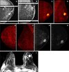
Images obtained in a 50-year-old female patient with a new
biopsy-proven malignant lesion in the left breast. (A) Craniocaudal
mammogram of the right breast does not show any lesion. (B) The malignant
lesion corresponds with a 7.0-cm irregular and spiculated mass on the left
craniocaudal mammogram. US-guided core-needle biopsy revealed grade 2
invasive lobular carcinoma. (C) The bilateral positron emission mammographic
craniocaudal color image obtained 1 hour after intravenous injection of 185
MBq of fluorine 18–labeled fluorodeoxyglucose (18F-FDG) shows a mass
with intense uptake in the left breast with known cancer and no abnormal
uptake in the right breast. Positron emission mammographic craniocaudal
images of the left breast obtained (D) 1 hour and (E) 4 hours after
intravenous injection of 185 MBq of 18F-FDG show no substantial visual
difference in uptake of the known cancer. (F) Axial contrast-enhanced
fat-saturated subtracted T1-weighted MR image with maximum intensity
projection reconstruction obtained 90 seconds after intravenous injection of
0.1 mmol of gadolinium-based contrast material per kilogram of body weight
also shows the enhancing mass corresponding to known malignancy (arrow) and
marked bilateral background parenchymal enhancement, with multiple
nonspecific foci of enhancement in the contralateral breast. The patient
opted for bilateral mastectomy, which confirmed left-sided malignancy and no
malignancy in the contralateral breast.
Figure 3:
Images obtained in a 50-year-old female patient with a new biopsy-proven malignant lesion in the left breast. (A) Craniocaudal mammogram of the right breast does not show any lesion. (B) The malignant lesion corresponds with a 7.0-cm irregular and spiculated mass on the left craniocaudal mammogram. US-guided core-needle biopsy revealed grade 2 invasive lobular carcinoma. (C) The bilateral positron emission mammographic craniocaudal color image obtained 1 hour after intravenous injection of 185 MBq of fluorine 18–labeled fluorodeoxyglucose (18F-FDG) shows a mass with intense uptake in the left breast with known cancer and no abnormal uptake in the right breast. Positron emission mammographic craniocaudal images of the left breast obtained (D) 1 hour and (E) 4 hours after intravenous injection of 185 MBq of 18F-FDG show no substantial visual difference in uptake of the known cancer. (F) Axial contrast-enhanced fat-saturated subtracted T1-weighted MR image with maximum intensity projection reconstruction obtained 90 seconds after intravenous injection of 0.1 mmol of gadolinium-based contrast material per kilogram of body weight also shows the enhancing mass corresponding to known malignancy (arrow) and marked bilateral background parenchymal enhancement, with multiple nonspecific foci of enhancement in the contralateral breast. The patient opted for bilateral mastectomy, which confirmed left-sided malignancy and no malignancy in the contralateral breast.
Images obtained in an 85-year-old female patient with a new
biopsy-proven malignant lesion in the right breast. (A) The malignant lesion
corresponds with a 3.0-cm equal-density mildly irregular mass on right
craniocaudal mammogram. US-guided core-needle biopsy revealed grade 3
invasive intracystic papillary carcinoma. (B) Right positron emission
mammographic (PEM) craniocaudal color image obtained 1 hour after
intravenous injection of 74 MBq of fluorine 18–labeled
fluorodeoxyglucose (18F-FDG) shows a 3.2-cm mass (known cancer) with intense
uptake. Left PEM (C) mediolateral oblique and (D) craniocaudal color images
obtained 1 hour after intravenous injection of 74 MBq of 18F-FDG do not show
any abnormal uptake. Right PEM craniocaudal images obtained (E) 1 hour and
(F) 4 hours after intravenous injection of 74 MBq of 18F-FDG show no
substantial visual difference in uptake of the known cancer. (G, H) Axial
contrast-enhanced fat-saturated subtracted T1-weighted MR images with
maximum intensity projection reconstruction obtained 90 seconds after
intravenous injection of 0.1 mmol of gadolinium-based contrast material per
kilogram of body weight also show the enhancing mass corresponding to known
malignancy and multiple nonspecific foci of enhancement in the contralateral
breast, the largest in the left central breast with washout kinetics (arrow
in H). Left-sided MRI-guided biopsy confirmed benign intraductal papilloma.
The patient underwent a right lumpectomy with sentinel lymph node biopsy.
The final pathologic result confirmed 3.2-cm grade 3 invasive intracystic
papillary carcinoma with negative sentinel lymph node biopsy.
Figure 4:
Images obtained in an 85-year-old female patient with a new biopsy-proven malignant lesion in the right breast. (A) The malignant lesion corresponds with a 3.0-cm equal-density mildly irregular mass on right craniocaudal mammogram. US-guided core-needle biopsy revealed grade 3 invasive intracystic papillary carcinoma. (B) Right positron emission mammographic (PEM) craniocaudal color image obtained 1 hour after intravenous injection of 74 MBq of fluorine 18–labeled fluorodeoxyglucose (18F-FDG) shows a 3.2-cm mass (known cancer) with intense uptake. Left PEM (C) mediolateral oblique and (D) craniocaudal color images obtained 1 hour after intravenous injection of 74 MBq of 18F-FDG do not show any abnormal uptake. Right PEM craniocaudal images obtained (E) 1 hour and (F) 4 hours after intravenous injection of 74 MBq of 18F-FDG show no substantial visual difference in uptake of the known cancer. (G, H) Axial contrast-enhanced fat-saturated subtracted T1-weighted MR images with maximum intensity projection reconstruction obtained 90 seconds after intravenous injection of 0.1 mmol of gadolinium-based contrast material per kilogram of body weight also show the enhancing mass corresponding to known malignancy and multiple nonspecific foci of enhancement in the contralateral breast, the largest in the left central breast with washout kinetics (arrow in H). Left-sided MRI-guided biopsy confirmed benign intraductal papilloma. The patient underwent a right lumpectomy with sentinel lymph node biopsy. The final pathologic result confirmed 3.2-cm grade 3 invasive intracystic papillary carcinoma with negative sentinel lymph node biopsy.
Images obtained in a 69-year-old female patient with a new
biopsy-proven malignant lesion in the left breast. (A, B) The malignant
lesion corresponds with a 2.5-cm irregular and spiculated mass (arrow) and
an additional oval and circumscribed mass (double arrows) on left
mediolateral oblique and craniocaudal spot compression mammographic views.
US-guided core-needle biopsy revealed grade 3 invasive ductal carcinoma
(arrow) and fibroadenoma (double arrow). Left positron emission mammographic
(PEM) (C) craniocaudal and (D) mediolateral oblique color images obtained 1
hour after intravenous injection of 74 MBq of fluorine 18–labeled
fluorodeoxyglucose (18F-FDG) show a mass (known cancer) with intense uptake
and no uptake in the biopsy-proven fibroadenoma. Right PEM (E) craniocaudal
and (F) mediolateral oblique color images obtained 1 hour after intravenous
injection of 74 MBq of 18F-FDG do not show any abnormal uptake. Left PEM
craniocaudal images obtained (G) 1 hour and (H) 4 hours after intravenous
injection of 74 MBq of 18F-FDG show no substantial visual difference in
uptake of known cancer. (I) Axial contrast-enhanced fat-saturated subtracted
T1-weighted MR image with maximum intensity projection reconstruction
obtained 90 seconds after intravenous injection of 0.1 mmol of
gadolinium-based contrast material per kilogram of body weight also shows
the left-sided enhancing mass (arrow) corresponding to known malignancy and
the biopsy-proven fibroadenoma (double arrows), as well as multiple
nonspecific foci of enhancement in the contralateral breast, with one being
the most conspicuous (arrow). The patient underwent bilateral lumpectomy.
The final pathologic results in the left breast confirmed grade 3 invasive
ductal carcinoma and yielded atypical ductal hyperplasia in the right
breast.
Figure 5:
Images obtained in a 69-year-old female patient with a new biopsy-proven malignant lesion in the left breast. (A, B) The malignant lesion corresponds with a 2.5-cm irregular and spiculated mass (arrow) and an additional oval and circumscribed mass (double arrows) on left mediolateral oblique and craniocaudal spot compression mammographic views. US-guided core-needle biopsy revealed grade 3 invasive ductal carcinoma (arrow) and fibroadenoma (double arrow). Left positron emission mammographic (PEM) (C) craniocaudal and (D) mediolateral oblique color images obtained 1 hour after intravenous injection of 74 MBq of fluorine 18–labeled fluorodeoxyglucose (18F-FDG) show a mass (known cancer) with intense uptake and no uptake in the biopsy-proven fibroadenoma. Right PEM (E) craniocaudal and (F) mediolateral oblique color images obtained 1 hour after intravenous injection of 74 MBq of 18F-FDG do not show any abnormal uptake. Left PEM craniocaudal images obtained (G) 1 hour and (H) 4 hours after intravenous injection of 74 MBq of 18F-FDG show no substantial visual difference in uptake of known cancer. (I) Axial contrast-enhanced fat-saturated subtracted T1-weighted MR image with maximum intensity projection reconstruction obtained 90 seconds after intravenous injection of 0.1 mmol of gadolinium-based contrast material per kilogram of body weight also shows the left-sided enhancing mass (arrow) corresponding to known malignancy and the biopsy-proven fibroadenoma (double arrows), as well as multiple nonspecific foci of enhancement in the contralateral breast, with one being the most conspicuous (arrow). The patient underwent bilateral lumpectomy. The final pathologic results in the left breast confirmed grade 3 invasive ductal carcinoma and yielded atypical ductal hyperplasia in the right breast.

References

    1. Allgood PC , Duffy SW , Kearins O , et al. . Explaining the difference in prognosis between screen-detected and symptomatic breast cancers . Br J Cancer 2011. ; 104 ( 11 ): 1680 – 1685 . - PMC - PubMed
    1. Smith RA , Duffy SW , Gabe R , Tabar L , Yen AM , Chen TH . The randomized trials of breast cancer screening: what have we learned? Radiol Clin North Am 2004. ; 42 ( 5 ): 793 – 806 , v. - PubMed
    1. Boyd NF , Martin LJ , Yaffe MJ , Minkin S . Mammographic density and breast cancer risk: current understanding and future prospects . Breast Cancer Res 2011. ; 13 ( 6 ): 223 . - PMC - PubMed
    1. Kerlikowske K , Zhu W , Tosteson AN , et al. . Identifying women with dense breasts at high risk for interval cancer: a cohort study . Ann Intern Med 2015. ; 162 ( 10 ): 673 – 681 . - PMC - PubMed
    1. Kuhl CK , Strobel K , Bieling H , Leutner C , Schild HH , Schrading S . Supplemental Breast MR Imaging Screening of Women with Average Risk of Breast Cancer . Radiology 2017. ; 283 ( 2 ): 361 – 370 . - PubMed

Substances

Associated data