. 2024 Jan;13(2):e6984.
doi: 10.1002/cam4.6984.

PIEZO1 is essential for the survival and proliferation of acute myeloid leukemia cells

Affiliations

PIEZO1 is essential for the survival and proliferation of acute myeloid leukemia cells

Delphine Lebon et al. Cancer Med. 2024 Jan.

Abstract

Introduction: Leukemogenesis is a complex process that interconnects tumoral cells with their microenvironment, but the effect of mechanosensing in acute myeloid leukemia (AML) blasts is poorly known. PIEZO1 perceives and transmits the constraints of the environment to human cells by acting as a non-selective calcium channel, but very little is known about its role in leukemogenesis.

Results: For the first time, we show that PIEZO1 is preferentially expressed in healthy hematopoietic stem and progenitor cells in human hematopoiesis, and globally overexpressed in AML cells. In AML subtypes, PIEZO1 expression associates with favorable outcomes as better overall (OS) and disease-free survival (DFS). If PIEZO1 is expressed and functional in THP1 leukemic myeloid cell line, its chemical activation doesn't impact the proliferation, differentiation, nor survival of cells. However, the downregulation of PIEZO1 expression dramatically reduces the proliferation and the survival of THP1 cells. We show that PIEZO1 knock-down blocks the cell cycle in G0/G1 phases of AML cells, impairs the DNA damage response pathways, and critically increases cell death by triggering extrinsic apoptosis pathways.

Conclusions: Altogether, our results reveal a new role for PIEZO1 mechanosensing in the survival and proliferation of leukemic blasts, which could pave the way for new therapeutic strategies to target AML cells.

Keywords: PIEZO1; acute myeloid leukemia; apoptosis; cell cycle; proliferation; survival.


Conflict of interest statement

The authors declare no competing financial interests.

Figures
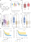
FIGURE 1
FIGURE 1
PIEZO1 expression pattern in normal hematopoietic cells and prognostic value in AML cohorts (MILE and TCGA). (A) PIEZO1 mRNA expression was determined by quantitative reverse transcriptase‐polymerase chain reaction (RT‐qPCR) relative to GAPDH expression. Different cell fractions were sorted from peripheral hematopoietic stem cells and bone marrow as described in the methods section. n = 5; *** p < 0.001. (B) PIEZO1 mRNA expression in 352 AML with normal karyotype and other abnormalities, 107 AML with favorable cytogenetic, 38 AML with 11q23 rearrangement, and 48 AML with complex karyotype compare to 73 healthy bone marrow (MILE GSE13159 cohort). (C) TCGA database (200 AML patients for whom genome or whole exome analysis was available) analysis of PIEZO1 mRNA expression showed an increased bone marrow blast rate for patients PIEZO1 expression over than median (p = 0.016). (D) TCGA database analysis of PIEZO1 mRNA expression (Z‐score) according to cytological AML subtypes M1 (M3 subtypes were excluded) (p = 0.00057). (E) TCGA database analysis of PIEZO1 mRNA expression (Z‐score) according to cytogenetics subgroups as defined by ELN 2017 classification (0.81 ± 1.09 vs. −0.21 ± 0.92 and −0.12 ± 0.86 for, respectively, intermediate and adverse cytogenetic, p = 5.3 × 10−6). (F) TCGA database analysis of PIEZO1 mRNA expression (Z‐score) according to molecular profiles as defined by ELN 2017 classification (0.83 ± 1.07 vs. −0.23 ± 0.91 and −0.14 ± 0.88 for, respectively, intermediate and adverse molecular profiles, p = 1.5 × 10−6). (G) Overall survival of AML patients with high (higher than the third quartile) and low (below) PIEZO1 mRNA expression (p = 0.028, from TCGA database). (H) Disease‐free survival of AML patients with high (higher than the third quartile) and low (below) PIEZO1 mRNA expression (p = 0.01, from TCGA database). *p < 0.05; **p < 0.01; ***p < 0.001; ****p < 0.0001.
FIGURE 2
FIGURE 2
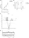
Effects of PIEZO1 KD on THP1 proliferation and transcriptomic data. All experiments were performed at less in triplicate; *** p < 0.001; ** p < 0.01; * p < 0.05. (A) THP1 total cell number at Day 7 in Scrambl shRNA (1,73 × 107 ± 1,82 × 107) and Sh PIEZO1 (3,61 × 105 ± 2,48 × 105). (B) Percentage of GFP+ cells at Day 4 (75.33% ± 16.04%) and Day 7 (23.57% ± 8.14%) in THP1 transduced with Sh PIEZO1 or with the Scrambl shRNA (92% ± 0.57 at Day 4 and XXXXX at Day 7). (C) Volcanoplot of differential gene expression between THP1 cells transduced with shPIEZO1 and Scrambl, panel of 770 genes from the panCancer pathway (NanoString Technologies). X‐axis is log2 (fold change), and Y‐axis is −log10 (adjusted p‐value). Significantly differentially expressed genes are indicated above the horizontal dashed line (adjusted p‐value = 0.01). (D) 26 genes were significantly deregulated (8 up‐ and 18 downregulated with a clear clustering between the two conditions) with an absolute value of Log2 (fold change) >2 and an adjusted p‐value <0.01.
FIGURE 3
FIGURE 3
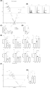
Effects of PIEZO1 KD on THP1 cell cycle and DNA damage pathway. All experiments were performed in triplicate; *** p < 0.001; ** p < 0.01; * p < 0.05. (A) Volcanoplot of expression of genes involved in cell cycle and apoptosis pathways deregulated after PIEZO1 KD, panel of 770 genes from the panCancer pathway (NanoString Technologies). X‐axis is log2 (fold change), and Y‐axis is −log10 (adjusted p‐value). (B) and (C) Cell cycle analysis of THP1 cells stained with Hoechst and analyzed by MFC revealed a blockage in G0/G1 phases (64.5% with Sh#1 and 57.4% with Sh#2 vs. 41% with Scrambl) and a significant decrease of G2/M cell cycle phases (13.8% with Sh#1 and 15.7% with Sh#2 vs. 28% with Scrambl) in PIEZO1 KD conditions. (D) MFC assessment of CD14 expression in THP1 cells 4 days after PIEZO1 KD (CD14 MFI Scrambl: 1.74 ± 0.6 and Sh PIEZO1: 7.12 ± 1.24). (E) Western blot analysis of key proteins of G0/G1 transition in THP1 cells, intensity normalized to beta‐actin and to Scrambl cells (Scr): cyclin D1 (Sh#1: 0.55 ± 0.31, Sh#2: 0.35 ± 0.26), cyclin E1 (Sh#1: 0.55 ± 0.21, Sh#2: 0.48 ± 0.25), CDC25A (Sh#1: 0.73 ± 0.13, Sh#2: 0.43 ± 0.12), CDK2 (Sh#1: 0.4 ± 0.25, Sh#2: 0.44 ± 0.1), CDK4 (Sh#1: 0.51 ± 0.11, Sh#2: 0.59 ± 0.09), and CDK6 (Sh#1: 0.74 ± 0.16, Sh#2: 0.53 ± 0.07) relative to β actin. Phospho Rb Ser‐780 (Sh#1: 0.65 ± 0.05, Sh#2: 0.53 ± 0.32) and phospho Rb Ser‐612 (Sh#1: 1.66 ± 0.38, Sh#2: 2.29 ± 0.36) were evaluated relative to Rb protein and to Scrambl cells. (F) Volcanoplot of expression of genes involved in DNA damage pathways deregulated after PIEZO1 KD. X‐axis is log2 (fold change), and Y‐axis is −log10 (adjusted p‐value). (G) Western blot of p‐γH2AX level in THP1 cells transduced with shPIEZO1 (3.895 ± 1.04 with Sh#1 and 4.53 ± 1.87 with Sh#2), normalized to beta‐actin and compared to the Scrambl.
FIGURE 4
FIGURE 4
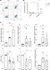
Effects of PIEZO1 KD on THP1 apoptosis. All experiments were performed in triplicate; *** p < 0.001; ** p < 0.01; * p < 0.05. (A) Assessment of Annexin V positive cells by MFC after PIEZO1 KD (17% ± 6.56 in shPIEZO1 vs. 1% in Scrambl cells). (B) Reversion assay of apoptosis adding 20 μM of the pan‐caspase inhibitor QVD in the medium at Day 4 post‐infection (7.67% ± 4.04 with shPIEZO1 vs. 1% with Scrambl). (C) Western blot analysis of proteins involved in apoptosis: PARP (Scrambl: 0.29 ± 0.03, Sh#1: 0.95 ± 0.04, SH#2: 1.89 ± 0.37), caspase 8 (Sh#1: 7.84 ± 7.1, Sh#2: 4.5 ± 3.1), caspase 3 (Sh#1: 15.3 ± 12.2, Sh#2: 12.3 ± 8.7), and caspase 9 (Sh#1: 3.25 ± 1.43, Sh#2: 2.38 ± 1.7) relative to their non‐cleaved forms, BCL‐2 (Sh#1: 0.6 ± 0.14, Sh#2: 0.5 ± 0.12) and c‐FLIP (Sh#1: 0.51 ± 0.26, Sh#2: 0.67 ± 0.2) relative to β actin.

References

    1. Lane SW, Gilliland DG. Leukemia stem cells. Semin Cancer Biol. 2010;20(2):71‐76. doi:10.1016/j.semcancer.2009.12.001 - DOI - PubMed
    1. Gilliland DG. Molecular genetics of human leukemias: new insights into therapy. Semin Hematol. 2002;39(4 Suppl 3):6‐11. doi:10.1053/shem.2002.36921 - DOI - PubMed
    1. Shlush LI, Zandi S, Mitchell A, et al. Identification of pre‐leukaemic haematopoietic stem cells in acute leukaemia. Nature. 2014;506(7488):328‐333. doi:10.1038/nature13038 - DOI - PMC - PubMed
    1. Corces‐Zimmerman MR, Hong WJ, Weissman IL, Medeiros BC, Majeti R. Preleukemic mutations in human acute myeloid leukemia affect epigenetic regulators and persist in remission. Proc Natl Acad Sci USA. 2014;111(7):2548‐2553. doi:10.1073/pnas.1324297111 - DOI - PMC - PubMed
    1. Bonnet D. Normal and leukaemic stem cells. Br J Haematol. 2005;130(4):469‐479. doi:10.1111/j.1365-2141.2005.05596.x - DOI - PubMed

Publication types