. 2024 Feb 5;14(3):319.
doi: 10.3390/nano14030319.

Imaging of Endometriotic Lesions Using cRGD-MN Probe in a Mouse Model of Endometriosis

Affiliations

Imaging of Endometriotic Lesions Using cRGD-MN Probe in a Mouse Model of Endometriosis

Nazanin Talebloo et al. Nanomaterials (Basel). .

Abstract

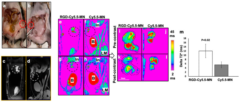
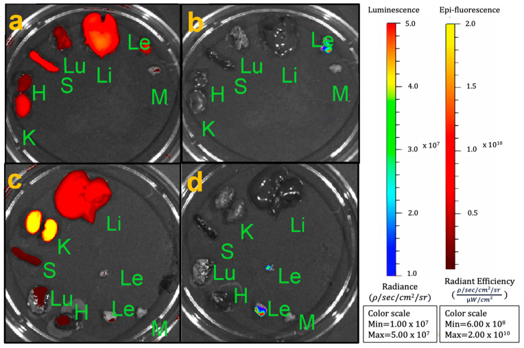
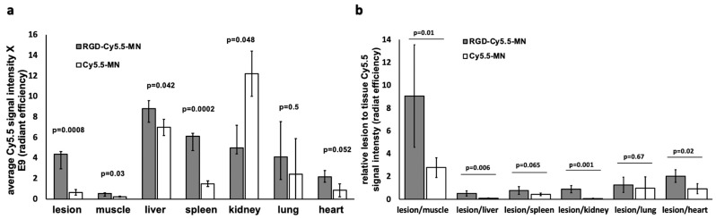
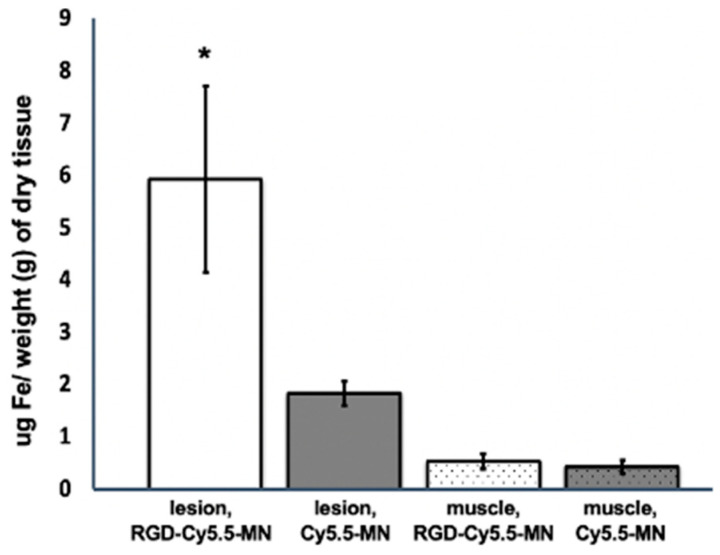
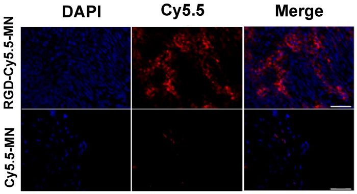
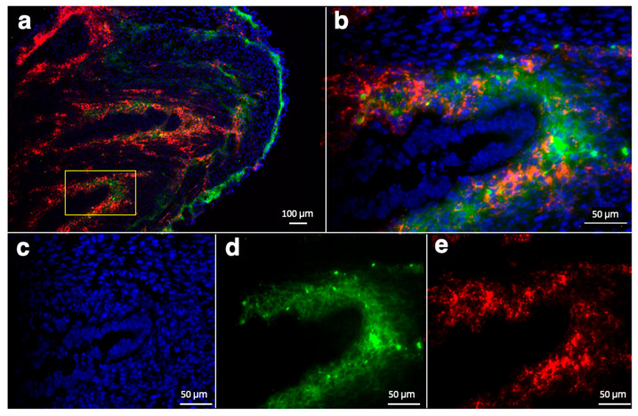
Approximately 10% of women suffer from endometriosis during their reproductive years. This disease is a chronic debilitating condition whose etiology for lesion implantation and survival heavily relies on adhesion and angiogenic factors. Currently, there are no clinically approved agents for its detection. In this study, we evaluated cRGD-peptide-conjugated nanoparticles (RGD-Cy5.5-MN) to detect lesions using magnetic resonance imaging (MRI) in a mouse model of endometriosis. We utilized a luciferase-expressing murine suture model of endometriosis. Imaging was performed before and after 24 h following the intravenous injection of RGD-Cy5.5-MN or control nanoparticles (Cy5.5-MN). Next, we performed biodistribution of RGD-Cy5.5-MN and correlative fluorescence microscopy of lesions stained for CD34. Tissue iron content was determined using inductively coupled plasma optical emission spectrometry (ICP-OES). Our results demonstrated that targeting endometriotic lesions with RGD-Cy5.5-MN resulted in a significantly higher delta T2* upon its accumulation compared to Cy5.5-MN. ICP-OES showed significantly higher iron content in the lesions of the animals in the experimental group compared to the lesions of the animals in the control group. Histology showed colocalization of Cy5.5 signal from RGD-Cy5.5-MN with CD34 in the lesions pointing to the targeted nature of the probe. This work offers initial proof-of-concept for targeting angiogenesis in endometriosis which can be useful for potential clinical diagnostic and therapeutic approaches for treating this disease.

Keywords: contrast agent; endometriosis; integrins; lesions; magnetic resonance imaging; nanoparticle.


Conflict of interest statement

The authors declare no conflicts of interest.

Figures

Figure 1
Figure 1
Representative photographs and T2* maps of the lesions in a mouse model of endometriosis. (a,b) Representative ex vivo photographs of endometriotic lesions shown within the red dotted circles. (c,d) Representative pre-contrast T2* maps (black and white color-coded) with endometriotic lesions shown within white dotted circles. (e) Pre-contrast and (g) post-contrast T2* maps of animals injected with RGD-Cy5.5-MN. (f) Pre-contrast and (h) post-contrast T2* maps of animals injected with Cy5.5-MN. (i) Magnified T2* maps of the lesions before and (k) after RGD-Cy5.5-MN administration. (j) Magnified T2* maps of the lesions captured before and (l) after Cy5.5-MN administration. Dotted area shows borders of the lesions. (m) Quantitative data (delta T2*) from the images (T2* maps) in (il). B—bladder; In—intestine; LM—leg muscle. Endometriotic lesions are shown within blue dotted circles in (ei,k).
Figure 2
Figure 2
Ex vivo biodistribution studies. (a,c) Fluorescence imaging of the biodistribution assessment of (a) experimental RGD-Cy5.5-MN and (c) control Cy5.5-MN probes; (b,d) Bioluminescence imaging showing the signal coming only from luciferase-expressing lesions in experimental and control groups. K—kidney; H—heart, S—spleen, Lu—lungs; Li—liver; Le—lesion; M—muscle.
Figure 3
Figure 3
(a) Quantification of ex vivo biodistribution shows significantly higher lesion accumulation of RGD-Cy5.5-MN compared to the control group. (b) Cy5.5 signal of the lesion normalized to the signal from other organs shows a significantly higher ratio when normalized to muscle, kidney, and heart.
Figure 4
Figure 4
ICP-OES analysis of the average iron content in endometriotic lesions and muscle tissues after intravenous administration of experimental (RGD-Cy5.5-MN) or control (Cy5.5-MN) probe. The iron content within the tissues was normalized to the dry weight of the tissues. Results are presented as means ± SD. Note that the lesions in animals injected with the experimental probe showed a higher iron content compared to that in animals injected with the control probe. The asterisk signifies a statistical significance (p < 0.01) in the comparison of iron content between lesions in the experimental and control groups.
Figure 5
Figure 5
Fluorescence microscopy demonstrates a relatively higher accumulation of RGD-Cy5.5-MN (top row) in endometriotic lesions compared to Cy5.5-MN (bottom row). Blue—DAPI nuclear stain; red—Cy5.5. Bar = 50 µm.
Figure 6
Figure 6
Immunofluorescence staining of endometriotic lesion sections from animals injected with RGD-Cy5.5-MN shown with (a) low; bar = 100 μm and (b) high; bar = 50 μm magnification. Note that (b) shows the area within the yellow rectangle in (a). Cell nucleus DAPI staining (c), CD34 marker (d, green) and Cy5.5 (e, red) are shown separately in the bottom row.

Similar articles

References

    1. Malvezzi H., Marengo E.B., Podgaec S., Piccinato C.d.A. Endometriosis: Current challenges in modeling a multifactorial disease of unknown etiology. J. Transl. Med. 2020;18:311. doi: 10.1186/s12967-020-02471-0. - DOI - PMC - PubMed
    1. Zondervan K.T., Becker C.M., Koga K., Missmer S.A., Taylor R.N., Viganò P. Endometriosis. Nat. Rev. Dis. Primers. 2018;4:9. doi: 10.1038/s41572-018-0008-5. - DOI - PubMed
    1. Kodaman P.H. Current strategies for endometriosis management. Obstet. Gynecol. Clin. 2015;42:87–101. doi: 10.1016/j.ogc.2014.10.005. - DOI - PubMed
    1. Prentice A. Endometriosis. BMJ. 2001;323:93–95. doi: 10.1136/bmj.323.7304.93. - DOI - PMC - PubMed
    1. Agarwal S.K., Chapron C., Giudice L.C., Laufer M.R., Leyland N., Missmer S.A., Singh S.S., Taylor H.S. Clinical diagnosis of endometriosis: A call to action. Am. J. Obstet. Gynecol. 2019;220:354.e1–354.e12. doi: 10.1016/j.ajog.2018.12.039. - DOI - PubMed

LinkOut - more resources