. 2024 Jan 23;13(3):204.
doi: 10.3390/cells13030204.

Prospective Approach to Deciphering the Impact of Intercellular Mitochondrial Transfer from Human Neural Stem Cells and Brain Tumor-Initiating Cells to Neighboring Astrocytes

Affiliations

Prospective Approach to Deciphering the Impact of Intercellular Mitochondrial Transfer from Human Neural Stem Cells and Brain Tumor-Initiating Cells to Neighboring Astrocytes

Jerusha Boyineni et al. Cells. .

Abstract

The communication between neural stem cells (NSCs) and surrounding astrocytes is essential for the homeostasis of the NSC niche. Intercellular mitochondrial transfer, a unique communication system that utilizes the formation of tunneling nanotubes for targeted mitochondrial transfer between donor and recipient cells, has recently been identified in a wide range of cell types. Intercellular mitochondrial transfer has also been observed between different types of cancer stem cells (CSCs) and their neighboring cells, including brain CSCs and astrocytes. CSC mitochondrial transfer significantly enhances overall tumor progression by reprogramming neighboring cells. Despite the urgent need to investigate this newly identified phenomenon, mitochondrial transfer in the central nervous system remains largely uncharacterized. In this study, we found evidence of intercellular mitochondrial transfer from human NSCs and from brain CSCs, also known as brain tumor-initiating cells (BTICs), to astrocytes in co-culture experiments. Both NSC and BTIC mitochondria triggered similar transcriptome changes upon transplantation into the recipient astrocytes. In contrast to NSCs, the transplanted mitochondria from BTICs had a significant proliferative effect on the recipient astrocytes. This study forms the basis for mechanistically deciphering the impact of intercellular mitochondrial transfer on recipient astrocytes, which will potentially provide us with new insights into the mechanisms of mitochondrial retrograde signaling.

Keywords: astrocytes; cancer stem cells; intercellular mitochondrial transfer; neural stem cells.


Conflict of interest statement

The authors declare no conflict of interest.

Figures

Figure 1
Figure 1
(a) Immunofluorescence analysis of astrocytes. Differentiated astrocytes (Ast) and LC26-10R (RG) were stained with antibodies against astrocyte differentiation markers, CD44, EAAT2, GFAP, Nestin (NES), SOX2, and Vimentin (VIM). Nuclei were counterstained with DAPI (blue). (b) RT-PCR analysis for astrocyte differentiation. Gene expression levels of stemness marker, BLBP, and astrocyte marker EAAT2 in Type 1 (Ast 1) and Type 2 (Ast 2) astrocytes are shown.
Figure 2
Figure 2
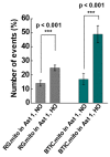
Intercellular mitochondrial transfer. To evaluate the direction of mitochondrial transfer from one cell type to the other in normoxic (NO) and hypoxic (HO) conditions, a co-culture was set up with 2 cell types by staining one cell population with MitoTracker (Mito.Red) and the other left unstained. Sets of cell types used for co-culturing were LC26-10R (RG) [13] (Mito.Red) + Ast.Type1, and LC26-RTL (BTIC) [13] (Mito.Red) + Ast.Type1. The graphs represent the average number of unstained cells that received mitochondria per 100 unstained cells. A 2x2 ANOVA (oxygenation by cell type) revealed the main effect of oxygenation (F(1,48) = 31.8, p < 0.001), the main effect of cell type (F (1,48) = 11.85, p = 0.001), and a significant interaction between oxygenation and cell type (F(1,48) = 6.59, p = 0.013). BTICs and RG cells showed significant increases in mitochondrial transfer in HO compared with NO (BTIC: t(23) = 4.33, p < 0.001; RG: t(25) = 3.85, p < 0.001). The graphs represent the average number of unstained cells that received mitochondria per 100 unstained cells. *** p < 0.001.
Figure 3
Figure 3
(a) TNT formation in LC26-10R (RG) and LC26-RTL (BTIC) cells compared in normoxic and hypoxic conditions. TNTs indicated with arrows. (b) Mitochondrial transfer within the same cell type in normoxic (NO) and hypoxic (HO) conditions. A co-culture was set up with half of the cell population stained with MitoTracker (Mito.Red) and the other half left unstained. The mitochondrial transfer among LC26-10R or LC26-RTL cells [13] was compared in normoxia and hypoxia. The graphs represent the percentage of unstained cells that received mitochondria per 100 unstained cells. A 2 × 2 ANOVA of cell type and oxygenation revealed the main effect of cell type (F(1,41) = 8.19, p = 0.007), the main effect of oxygenation (F(1,41) = 24.24, p < 0.001), and a significant interaction between cell type and oxygenation (F(1,41) = 8.19, p = 0.007). Post hoc tests showed a significant difference in mitochondrial transfer in BTICs as a function of oxygenation (t(17) = 5.71, p < 0.001) and a trending difference for RG cells (t(24) = 1.96, p = 0.06). *** p < 0.001.
Figure 4
Figure 4
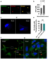
(a) Extracted mitochondria stained with MitoTracker Red and MitoTracker Green. (b) ATP quantity was measured in increasing amounts of mitochondrial extracts using a luciferase assay. (c) Mitochondria (red) from LC26-10R (RG) and (d) LC26-RTL (BTIC) transplanted into astrocytes. (e) Transplantation efficiency: The graph represents the average number of cells with transplanted mitochondria per 100 cells. (f) Mitochondria (red) transferred from LC26-RTL (BTIC) to LC26-10R (RG) through TNT in a co-culture experiment. Nanotube with mitochondria is magnified. (g) LC26-10R (RG) (cytoplasm in green; nucleus in blue) with transplanted mitochondria from LC26-RTL (BTIC) [13] in red. Nanotube with mitochondria is magnified.
Figure 5
Figure 5
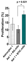
Cell proliferation. The percentage of cell proliferation in Type 1 astrocytes, Type 1 astrocytes with transplanted mitochondria from LC26-10R (RG), and Type 1 astrocytes with transplanted mitochondria from LC26-RTL (BTIC) [13]. A one-way ANOVA revealed the main effect of type (F (2,72) = 5.17, p = 0.008), with post hoc tests showing a significant difference in proliferation between Type 1 astrocytes, transplanted with RG mitochondria vs. Type 1 astrocytes, transplanted with BTIC mitochondria (p = 0.021). * p < 0.05.
Figure 6
Figure 6
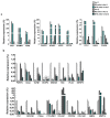
RT-PCR analysis for relative gene expression levels of differentially expressed genes between astrocytes (Types 1, 2) and astrocytes (Types 1, 2) with transplanted mitochondria from LC26-10R (RG) or LC26-RTL (BTIC) [13]. (a) Predominantly upregulated genes in astrocytes (Type 1) with transplanted mitochondria from LC26-10R(RG) or LC26-RTL (BTIC); (b) predominantly downregulated genes in astrocytes (Type 1) with transplanted mitochondria from LC26-10R (RG) or LC26-RTL (BTIC).
Figure 7
Figure 7
(a) Relative gene expression level of SOX2 in LC26-10R and LC26-10R(Dif). (b) Relative gene expression levels of SOX2, BLBP, cMyc, CD44, Caveolin 1, and Emp1 in differentiated LC26-10R (RG(Dif)), differentiated LC26-10R (RG(Dif)) with transplanted mitochondria from LC26-10R (RG), and differentiated LC26-10R (RG(Dif)) with transplanted mitochondria from LC26-RTL (BTIC) [13].

Similar articles

Cited by

References

    1. Valdebenito S., Audia A., Bhat K.P., Okafo G., Eugenin E.A. Tunneling Nanotubes Mediate Adaptation of Glioblastoma Cells to Temozolomide and Ionizing Radiation Treatment. iScience. 2020;23:101450. doi: 10.1016/j.isci.2020.101450. - DOI - PMC - PubMed
    1. Valdebenito S., Lou E., Baldoni J., Okafo G., Eugenin E. The Novel Roles of Connexin Channels and Tunneling Nanotubes in Cancer Pathogenesis. Int. J. Mol. Sci. 2018;19:1270. doi: 10.3390/ijms19051270. - DOI - PMC - PubMed
    1. Pinto G., Saenz-De-Santa-Maria I., Chastagner P., Perthame E., Delmas C., Toulas C., Moyal-Jonathan-Cohen E., Brou C., Zurzolo C. Patient-derived glioblastoma stem cells transfer mitochondria through tunneling nanotubes in tumor organoids. Biochem. J. 2021;478:21–39. doi: 10.1042/BCJ20200710. - DOI - PMC - PubMed
    1. Cuyàs E., Verdura S., Folguera-Blasco N., Bastidas-Velez C., Martin G., Alarcón T., Menendez J.A. Mitostemness. Cell Cycle. 2018;17:918–926. doi: 10.1080/15384101.2018.1467679. - DOI - PMC - PubMed
    1. Ferrón S.R., Charalambous M., Radford E., McEwen K., Wildner H., Hind E., Morante-Redolat J.M., Laborda J., Guillemot F., Bauer S.R., et al. Postnatal loss of Dlk1 imprinting in stem cells and niche astrocytes regulates neurogenesis. Nature. 2011;475:381–385. doi: 10.1038/nature10229. - DOI - PMC - PubMed

Publication types