. 2024 Jan 23;13(3):206.
doi: 10.3390/cells13030206.

Blastocyst-Derived Lactic Acid May Regulate S100A6 Expression and Function in Mouse Decidualization via Stimulation of Uterine Epithelial Arachidonic Acid Secretion

Affiliations

Blastocyst-Derived Lactic Acid May Regulate S100A6 Expression and Function in Mouse Decidualization via Stimulation of Uterine Epithelial Arachidonic Acid Secretion

Meng-Yuan Li et al. Cells. .

Abstract

(1) Background: Inflammatory responses are implicated in embryo implantation, decidualization, pregnancy maintenance and labor. Both embryo implantation and decidualization are essential to successful pregnancy in rodents and primates. S100A6 is involved in inflammation, tumor development, apoptosis and calcium homeostasis. S100A6 is strongly expressed in mouse decidua, but the underlying mechanisms of how S100A6 regulates implantation and decidualization are poorly defined. (2) Methods: Mouse endometrial stromal and epithelial cells are isolated from day 4 pseudopregnant mouse uteri. Both immunofluorescence and Western blotting are used to analyze the expression and localization of proteins. The molecular mechanism is verified in vitro by Western blotting and the quantitative polymerase chain reaction. (3) Results: From days 4 to 8 of pregnancy, S100A6 is specifically expressed in mouse subluminal stromal cells. Blastocyst-derived lactic acid induces AA secretion by activating the luminal epithelial p-cPLA2. The epithelial AA induces stromal S100A6 expression through the COX2/PGI2/PPAR δ pathway. Progesterone regulates S100A6 expression through the progesterone receptor (PR). S100A6/RAGE signaling can regulate decidualization via EGFR/ERK1/2 in vitro. (4) Conclusions: S100A6, as an inflammatory mediator, is important for mouse implantation and decidualization.

Keywords: RAGE; S100A6; decidualization; implantation; inflammation.


Conflict of interest statement

The authors declare no conflicts of interest.

Figures
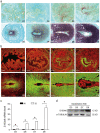
Figure 1
Figure 1
S100A6 expression in early pregnant mouse uteri. (a) S100A6 mRNA in situ hybridization from days 1 to 8 of pregnancy. (b) Immunofluorescence of S100A6 protein (green) and propidium iodide (PI; red) from days 1 to 8 of pregnancy. (c) RT-qPCR results of S100A6 mRNA levels at implantation sites (IS) and inter-implantation sites (NI) from days 5 to 8 of pregnancy. (d) Western blot results of S100A6 protein levels in mouse uteri at implantation sites (IS) from days 5 to 8 of pregnancy. Scale bar = 250 μm. D1-D8, days 1 to 8 of pregnancy. Bars represent mean ± SD; * p < 0.05.
Figure 2
Figure 2
Active blastocysts induce S100A6 expression. (a) Western blot results of S100A6 protein levels in day 4 pseudopregnant (PD4) and pregnant uteri (D4). (b) Immunofluorescence of S100A6 protein (green) and propidium iodide (PI; red) in day 4 pseudopregnant (PD4) and pregnant uteri (D4). (c) RT-qPCR results of uterine S100A6 mRNA levels undergoing delayed implantation and at implantation sites (IS) and inter-implantation sites (NI) of activated uteri. (d) In situ hybridization showing S100A6 expression undergoing delayed implantation and at the implantation site of an activated uterus. (e) Western blot analysis of S100A6 protein levels under delayed implantation and at implantation sites (IS) of activated uteri. (f) Immunofluorescence of S100A6 protein (green) and propidium iodide (PI, red) undergoing delayed implantation and at the implantation site of an activated uterus. Scale bar = 250 μm. Bars represent mean ± SD; * p < 0.05.
Figure 3
Figure 3
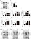
Blastocyst-derived lactic acid regulation of S100A6 expression is dependent on the AA/COX2/PGI2/PPAR δ pathway. (a) Western blot showing p-cPLA2 protein level in lactic acid-treated endometrial epithelial cells for 3 h. (b) ELISA analysis of the concentration of AA in the epithelial cell-culture medium. (c) Western blot showing S100A6, COX2, PGIs and PPAR δ protein levels in endometrial stromal cells treated with AA, AA and NS398, or NS398 for 3 h. (d) Western blot analysis of S100A6, COX2, PGIs and PPAR δ protein levels in endometrial stromal cells treated with AA, AA and GSK0660, or GSK0660 for 3 h. (e) Western blot analysis of S100A6 and PPAR δ protein levels in endometrial stromal cells treated with Iloprost, Iloprost and GSK0660, or GSK0660 alone for 3 h. (f) Western blot showing S100A6 and PPAR δ protein levels in endometrial stromal cells treated with GW501516, GW501516 and GSK0660, or GSK0660 for 3 h. (g) Western blot showing S100A6, COX2, PGIs and PPAR δ protein levels in stromal cells after cocultures of uterine epithelial cells and stromal cells were treated with lactic acid, lactic acid and AZD3965, or AZD3965 for 3 h. Lac, lactic acid. Bars represent mean ± SD; * p < 0.05.
Figure 4
Figure 4
Effects of S100A6 on mouse in vitro decidualization. (a) Western blot showing S100A6 protein levels after stromal cells were induced to undergo in vitro decidualization for 48 h. (b) RT-qPCR results of S100A6 mRNA levels during in vitro decidualization from 24 to 72 h. (c) RT-qPCR results of the effects of S100A6 siRNAs on S100A6 mRNA levels in endometrial stromal cells. (d) RT-qPCR results of the effects of S100A6 siRNA expression on Prl8a2 mRNA levels in endometrial stromal cells undergoing in vitro decidualization at 48 h. (e) RT-qPCR results of the effects of S100A6 siRNA expression on S100A6 mRNA levels in endometrial stromal cells undergoing in vitro decidualization at 48 h. (f) Western blot results of the effects of S100A6 siRNA expression on S100A6, SNAIL, E-cadherin and ZO-1 protein levels undergoing in vitro decidualization at 48 h. (g) RT-qPCR results of the effects of S100A6 overexpression on S100A6 mRNA levels in endometrial stromal cells undergoing in vitro decidualization at 48 h. (h) RT-qPCR analysis of the effects of S100A6 overexpression on Prl8a2 mRNA levels in endometrial stromal cells under in vitro decidualization at 48 h. (i) Western blot results of the effects of S100A6 overexpression on S100A6, SNAIL, E-cadherin and ZO-1 protein levels undergoing in vitro decidualization at 48 h. Si S1006, S100A6 siRNA; S100A6 OE, S100A6 overexpression; EP, estrogen and progesterone treatment. Bars represent mean ± SD; * p < 0.05; ** p < 0.01; ns, not significant.
Figure 5
Figure 5
S100A6 regulates decidualization via RAGE. (a) RAGE immunofluorescence (green) in mouse uteri from days 1 to 8 of pregnancy. (b) Western blot results of RAGE protein levels in mouse uteri at implantation sites (IS) from days 5 to 8 of pregnancy. (c) RT-qPCR results of the effects of FPS-ZM1 on Prl8a2 mRNA levels in endometrial stromal cells after in vitro decidualization for 48 h. (d) Western blot results of the effects of S100A6 siRNA expression on RAGE protein levels at 48 h of in vitro decidualization. (e) Western blot results of the effects of S100A6 overexpression on RAGE protein levels at 48 h of in vitro decidualization. D1–D8, days 1 to 8 of pregnancy. Si S100A6, S100A6 siRNA; S100A6 OE, S100A6 overexpression; EP, estrogen and progesterone treatment. Scale bar = 100 μm. Bars represent mean ± SD; * p < 0.05; ** p < 0.01; ns, not significant.
Figure 6
Figure 6
S100A6 mediates decidualization via RAGE/EGFR/ERK1/2. (a) The effects of S100A6 siRNA expression on protein levels of EGFR, P-EGFR, T-ERK1/2, P-ERK1/2 and S100A6 in cells undergoing in vitro decidualization at 48 h. (b) Effects of S100A6 siRNA on S100A6 mRNA levels under in vitro decidualization conditions at 48 h. (c) Effects of FPS-ZM1 on Prl8a2 mRNA levels due to S100A6 overexpression under in vitro decidualization conditions at 48 h. (d) Effects of an EGFR inhibitor on Prl8a2 mRNA levels in S100A6-overexpressing cells undergoing in vitro decidualization at 48 h. (e) Effects of U0126 on Prl8a2 mRNA levels in S100A6-overexpressing cells undergoing in vitro decidualization at 48 h. (f) Effects of FPS-ZM1 on S100A6 mRNA levels in S100A6-overexpressing cells undergoing in vitro decidualization at 48 h. (g) Effects of an EGFR inhibitor on S100A6 mRNA levels when S100A6-overexpressing cells at 48 h of in vitro decidualization. (h) Effects of U0126 on S100A6 mRNA levels when S100A6 was overexpressed in cells undergoing in vitro decidualization at 48 h. (i) Effects of FPS-ZM1 on RAGE, EGFR, P-EGFR, T-ERK1/2, P-ERK1/2 and S100A6 protein levels in S100A6-overxpressing cells at 48 h of in vitro decidualization. (j) Effects of an EGFR inhibitor on RAGE, EGFR, P-EGFR, T-ERK1/2, P-ERK1/2 and S100A6 protein levels when S100A6 was overexpressed cells undergoing in vitro decidualization at 48 h. (k) Effect of U0126 on RAGE, EGFR, P-EGFR, T-ERK1/2, P-ERK1/2 and S100A6 protein levels when S100A6 was overexpressed in cells undergoing in vitro decidualization at 48 h. Si S1006, S100A6 siRNA; S100A6 OE, S100A6 overexpression; EGFR in, EGFR inhibitor; EP, estrogen and progesterone treatment. Bars represent mean ± SD; * p < 0.05; ** p < 0.01; ns, not significant.
Figure 7
Figure 7
Progesterone regulates S100A6 expression via the PR. (a) Western blot analysis of S100A6 protein levels in mouse uteri treated with oil, progesterone, or progesterone and RU486 for 24 h. (b) RT-qPCR to detect S100A6 mRNA levels in mouse uteri treated with oil, progesterone, or progesterone and RU486 for 24 h. (c) Immunofluorescence of S100A6 protein expression (green). Scale bar = 100 μm. Bars indicate mean ± SD; * p < 0.05.
Figure 8
Figure 8
Estrogen regulates S100A6 expression in an ERα-dependent manner. (a) RT-qPCR to detect S100A6 mRNA levels in mouse uteri treated with oil, E2, E2 and ICI, PPT, or DPN for 24 h. (b) S100A6 protein levels in mouse uteri treated with oil, E2, E2 and ICI, PPT, or DPN. (c) S100A6 immunofluorescence (green) in mouse uteri treated with oil, E2, E2 and ICI, or PPT. (d) RT-qPCR to detect S100A6 mRNA levels in wild type and ERαKO mouse uteri treated with oil or E2 for 24 h. (e) S100A6 protein levels in wild type and ERαKO mouse uteri treated with oil or E2 for 24 h. (f) S100A6 immunofluorescence in wild type and ERαKO mouse uteri treated with oil or E2 for 24 h. Scale bar = 100 μm. Bars indicate mean ± SD; * p < 0.05; ns, not significant.
Figure 9
Figure 9
Graphic summary of the regulation and action of S100A6 during mouse implantation and decidualization. Blastocyst-derived lactate regulates S100A6 expression through the cPLA2/AA/COX2/PGI2/PPAR δ pathway. S100A6 regulates decidualization via the RAGE/EGFR/ERK pathway.

Similar articles

Cited by

References

    1. Ochoa-Bernal M.A., Fazleabas A.T. Physiologic Events of Embryo Implantation and Decidualization in Human and Non-Human Primates. Int. J. Mol. Sci. 2020;21:1973. doi: 10.3390/ijms21061973. - DOI - PMC - PubMed
    1. Quenby S. Implantation involves an initial pro-inflammatory response. Fertil. Steril. 2020;114:288–289. doi: 10.1016/j.fertnstert.2020.05.039. - DOI - PubMed
    1. Mor G., Cardenas I. Inflammation and pregnancy: The role of the immune system at the implantation site. Ann. N. Y. Acad. Sci. 2011;1221:80–87. doi: 10.1111/j.1749-6632.2010.05938.x. - DOI - PMC - PubMed
    1. Erlebacher A. Immunology of the maternal-fetal interface. Annu. Rev. Immunol. 2013;31:387–411. doi: 10.1146/annurev-immunol-032712-100003. - DOI - PubMed
    1. Zhang T., Zhu W. Early transient suppression of immune checkpoint proteins T-cell immunoglobulin mucin-3 and programmed cell death-1 in peripheral blood lymphocytes after blastocyst transfer is associated with successful implantation. Fertil. Steril. 2020;114:426–435. doi: 10.1016/j.fertnstert.2019.12.022. - DOI - PubMed

Publication types

Substances