. 2024 Jan 23;13(3):209.
doi: 10.3390/cells13030209.

The Role of CCL24 in Primary Sclerosing Cholangitis: Bridging Patient Serum Proteomics to Preclinical Data

Affiliations

The Role of CCL24 in Primary Sclerosing Cholangitis: Bridging Patient Serum Proteomics to Preclinical Data

Raanan Greenman et al. Cells. .

Abstract

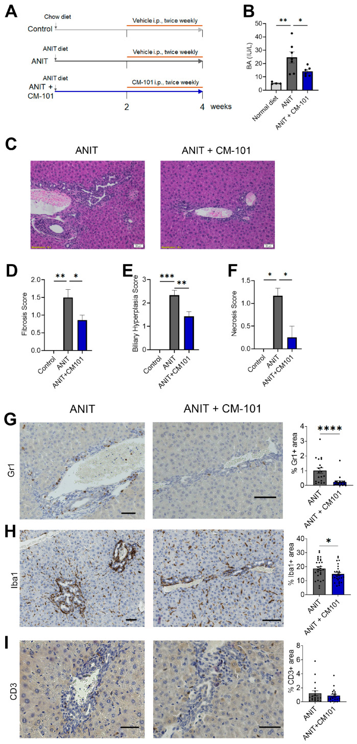
Primary sclerosing cholangitis (PSC) is an inflammatory and fibrotic biliary disease lacking approved treatment. We studied CCL24, a chemokine shown to be overexpressed in damaged bile ducts, and its involvement in key disease-related mechanisms. Serum proteomics of PSC patients and healthy controls (HC) were analyzed using the Olink® proximity extension assay and compared based on disease presence, fibrosis severity, and CCL24 levels. Disease-related canonical pathways, upstream regulators, and toxicity functions were elevated in PSC patients compared to HC and further elevated in patients with high CCL24 levels. In vitro, a protein signature in CCL24-treated hepatic stellate cells (HSCs) differentiated patients by disease severity. In mice, CCL24 intraperitoneal injection selectively recruited neutrophils and monocytes. Treatment with CM-101, a CCL24-neutralizing antibody, in an α-naphthylisothiocyanate (ANIT)-induced cholestasis mouse model effectively inhibited accumulation of peribiliary neutrophils and macrophages while reducing biliary hyperplasia and fibrosis. Furthermore, in PSC patients, CCL24 levels were correlated with upregulation of monocyte and neutrophil chemotaxis pathways. Collectively, these findings highlight the distinct role of CCL24 in PSC, influencing disease-related mechanisms, affecting immune cells trafficking and HSC activation. Its blockade with CM-101 reduces inflammation and fibrosis and positions CCL24 as a promising therapeutic target in PSC.

Keywords: CCL24; chemokines; cholangitis; fibrosis; hepatic stellate cells; inflammation; monocytes; neutrophils; proteomics.


Conflict of interest statement

Avi Katav, Raanan Greenman, Ilan Vaknin, Revital Aricha, Tom Snir, Matthew Frankel, John Lawler, and Adi Mor are employees of Chemomab Therapeutics (Israel). Massimo Pinzani is a Co-Founder and shareholder of Engitix Therapeutics Ltd. (UK), consultant of Chemomab, Chief Medical Advisor and shareholder of Aculive Therapeutics Ltd. (UK), Advisory board/consultant (active March 2022) of Chemomab Therapeutics (Israel); Resolution Therapeutics (UK); Dicerna (USA); Astra Zeneca (UK); and Galecto (Sweden). Amnon Peled is a consultant for Chemomab Therapeutics (Israel). Francesca Saffioti, Douglas Thorburn, Ophir Hay, and Inbal Mishalian have nothing to disclose.

Figures

Figure 1
Figure 1
PSC-related mechanisms are upregulated in patients with high CCL24 levels. (A) Analysis overview: sera from patients with PSC and HC were analyzed by the Olink Explore 3072 proteomic platform, and differentially expressed proteins (DEPs) were compared by disease, fibrosis, or CCL24. (B) Score plots of principal component analysis of proteome profiles in HC and patients with PSC with low or high ELF scores. (C) Correlation of CCL24 with representative proteins associated with inflammation/chemotaxis (CCL7 or CXCL10) in HC (n = 30) or in patients with PSC with ALP > 1.5 ULN (n = 34). (DI) Ingenuity pathway analysis of canonical pathways, upstream regulators, and liver-related toxicity functions. Venn diagrams show top 30 significant canonical pathways (D), top 20 significant upstream regulators (F), and significant liver-related toxicities (H). The average expression of protein lists of specific canonical pathways (E), upstream regulators (G), and liver-related toxicities (I) are presented for HC and patients with low and high CCL24 serum levels. Boxes represent interquartile ranges with medians (n = 20–30). * p < 0.05; ** p < 0.01; **** p < 0.0001. ELF, Enhanced Liver Fibrosis; HC, healthy controls; NA, not assigned; NPX, normalized protein expression.
Figure 2
Figure 2
PSC-related mechanisms are specifically associated with CCL24 and not with other eotaxins. (A) Correlation of CCL24, CCL11, and CCL26 in HC (n = 30) or patients with PSC with ALP > 1.5 ULN (n = 34). (B) The average expression of protein signatures was examined in patients with PSC, stratified by mean expression of each of the three eotaxins, CCL24, CCL11, and CCL26. Boxes represent interquartile ranges with medians (n = 20–25). * p < 0.05; ** p < 0.01, ns, no significance.
Figure 3
Figure 3
Hepatic cell line CCL24-dependent secreted proteins can differentiate individuals by PSC and its severity. (A) FACS analysis of LX-2 cells stained for CCR3. (B) Outline of the experimental procedure. LX2 cells were incubated with either PBS, CCL24, or CCL24 with CM-101. (C) Relative expression of the CCL24-dependent protein signature. (D) Differences in the serum proteomic profile of the CCL24-dependent signature were examined by an unsupervised heatmap of scaled expression values for each individual and identified protein. (E,F) The average expression of CCL24-dependent protein signatures is presented in HC vs. patients with PSC (E) or in patients with PSC, stratified by ELF score of 9.8 (F). Boxes represent interquartile ranges with medians (n = 20–30). (G,H) The ROC curves for the five serum proteins with the highest AUC values for predicting disease presence (G) or fibrosis severity (H). * p < 0.05; **** p < 0.0001.
Figure 4
Figure 4
CCL24 i.p. injection recruits neutrophils and monocytes. (A) Outline of the experimental procedure. (BF) Identification of peritoneal immune populations by scRNA-seq. (B) Uniform manifold approximation and projection (UMAP) plot of all cells that passed QC, clustered by the Louvain algorithm. (C) Heatmap of the top four gene markers for each cluster. Expression levels are scaled, and the number of cells in each column is downsampled to 500. (D) Stacked bar plot showing the relative abundance of each cell type, per treatment. (E) Violin plots of CCL24 expression levels by cell type. (F) Violin plots of CCL24 expression levels by cell type and treatment. (GJ) Identification of peritoneal immune populations by flow-cytometry. (G) Representative scatter plots of CD11b+ cells. (HJ) Quantification of relative change in cell counts for monocytes (H), neutrophils (I), and T cells (J). Data are represented as mean ± SEM (n = 4–10). * p < 0.05; ** p < 0.01.
Figure 5
Figure 5
CCL24 blockade attenuates neutrophil and macrophage accumulation and liver fibrosis in ANIT-induced cholestasis model. (A) Scheme of the experimental design. (B) Levels of serum bile acids (BA) under normal diet, ANIT diet, and ANIT diet with CM-101 (n = 4–7 mice). Data are represented as mean ± SEM. (C) Representative H&E staining in liver sections of ANIT diet or ANIT diet with CM-101. (DF) Histological scoring of fibrosis (D), biliary hyperplasia (E), and necrosis (F). Data are represented as mean ± SEM (n = 2–7). (GI) Representative immunohistochemistry staining and quantification of neutrophils (G), macrophages (H), and T cells (I). Data are represented as mean ± SEM (n = 4–7 mice, 4 fields per mouse). Scalebar represents 50 μm. * p < 0.05; ** p < 0.01; *** p < 0.001; **** p < 0.0001.
Figure 6
Figure 6
CCL24 is associated with monocyte and neutrophil migration in patients with PSC. (A,B) Serum proteomic profile of neutrophil chemotaxis and monocyte pathway gene lists were examined by a clustered heatmap of scaled expression values for each individual and protein. (CF) The average expression of protein lists of neutrophil chemotaxis (C), positive regulation of neutrophil migration (D), monocyte pathway (E), and positive regulation of monocyte chemotaxis (F) are presented for HC and patients with PSC, stratified by mean expression of each of the three eotaxins. Boxes represent interquartile ranges with medians (n = 20–30). * p < 0.05; ** p < 0.01; **** p < 0.0001, ns, no significance.

References

    1. Pollheimer M.J., Halilbasic E., Fickert P., Trauner M. Pathogenesis of Primary Sclerosing Cholangitis. Best Pract. Res. Clin. Gastroenterol. 2011;25:727–739. doi: 10.1016/j.bpg.2011.10.009. - DOI - PMC - PubMed
    1. Wu H., Chen C., Ziani S., Nelson L.J., Ávila M.A., Nevzorova Y.A., Cubero F.J. Fibrotic Events in the Progression of Cholestatic Liver Disease. Cells. 2021;10:1107. doi: 10.3390/cells10051107. - DOI - PMC - PubMed
    1. Poulsen K.L., Cajigas-Du Ross C.K., Chaney J.K., Nagy L.E. Role of the Chemokine System in Liver Fibrosis: A Narrative Review. Dig. Med. Res. 2022;5:30. doi: 10.21037/dmr-21-87. - DOI - PMC - PubMed
    1. Sahin H., Trautwein C., Wasmuth H.E. Functional Role of Chemokines in Liver Disease Models. Nat. Rev. Gastroenterol. Hepatol. 2010;7:682–690. doi: 10.1038/nrgastro.2010.168. - DOI - PubMed
    1. Braga T.T., Agudelo J.S.H., Camara N.O.S. Macrophages During the Fibrotic Process: M2 as Friend and Foe. Front. Immunol. 2015;6:602. doi: 10.3389/fimmu.2015.00602. - DOI - PMC - PubMed

Publication types

LinkOut - more resources