. 2024 Feb 1;65(2):21.
doi: 10.1167/iovs.65.2.21.

A Novel Exploration of the Choroidal Vortex Vein System: Incidence and Characteristics of Posterior Vortex Veins in Healthy Eyes

Affiliations

A Novel Exploration of the Choroidal Vortex Vein System: Incidence and Characteristics of Posterior Vortex Veins in Healthy Eyes

Guiqin He et al. Invest Ophthalmol Vis Sci. .

Abstract

Purpose: The purpose of this study was to investigate the incidence and characteristics of posterior vortex veins (PVVs) in healthy eyes and explore their relationship with age and refractive status.

Methods: This retrospective cross-sectional analysis encompassed 510 eyes from 255 consecutive healthy participants. Wide-field optical coherence tomography angiography (WF-OCTA) imaging was used to assess the presence of PVVs. Eyes were classified according to refractive status (emmetropia, low and moderate myopia, and high myopia) and age (minors and adults). The incidence and characteristics of eyes with PVVs were analyzed.

Results: Participants (mean age = 30.60 ± 21.12 years, 47.4% men) showed a mean refractive error of -2.83 ± 3.10 diopters (D; range = -12.00 to +0.75). PVVs were observed in 16.1% (82/510) of eyes. Of these, 39% (32/82) had PVVs in one eye and 61% (50/82) in both eyes. The mean number of PVVs per eye was 1.65 ± 1.05 (range = 1-6). PVVs are mainly around the optic disc (78%, 64/82) of eyes with PVVs and less in the macular area (6.1%, 5/82) or elsewhere (15.9%, 13/82). PVV incidence correlated with refractive status: 10.3% (22/213) in emmetropia, 16.6% (31/187) in low and moderate myopia, and 26.4% (29/110) in high myopia (P = 0.001), but not with age. Refractive status was the key predictor of PVV occurrence (odds ratio [OR] = 1.45, 95% confidence interval [CI] = 1.02-2.06, P = 0.038).

Conclusions: This study confirms PVVs' presence in healthy eyes, highlighting their inherent existence and susceptibility to alterations due to refractive conditions. These findings enhance our understanding of the vortex vein system and its distribution within the eyes.


Conflict of interest statement

Disclosure: G. He, None; X. Zhang, None; X. Zhuang, None; Y. Zeng, None; Y. Gan, None; Y. Su, None; M. Li, None; Y. Ji, None; L. Mi, None; X. Chen, None; F. Wen, None

Figures

Figure 1.
Figure 1.
Comparison of PVV detection using ICGA venous phase and en face WF-OCTA (Subjects not included). (A–E2) Two PVVs located around the optic disc in an eye with punctate inner choroidopathy. (A) Detection of two PVVs around the optic disc using the ICGA venous phase. (B) Detection of two PVVs around the optic disc using 24 × 20 mm en face WF-OCTA in the large-vessel choroidal layer. (C) WF-OCTA image showing the large-vessel choroidal layer in the same scan as B, with two horizontal lines (orange and blue arrows) crossing the exit points of PVVs (green arrowheads). (D) The yellow highlight indicates the presence of two PVVs around the optic disc in C. (E1) Horizontal WF-OCT image of C (orange arrow) corresponds to the 646th scan out of a total of 1280 WF-OCT scans, clearly demonstrating the exit of one PVV penetrating the sclera (green arrowhead) and leaving the eyeball. (E2) Horizontal WF-OCT image of C (blue arrow) corresponds to the 596th scan out of a total of 1280 WF-OCT scans, clearly illustrating the exit of another PVV penetrating the sclera (green arrowhead) and leaving the eyeball. (F–J2) Two PVVs located around the optic disc in an eye with acute zonal occult outer retinopathy. (F) Detection of two PVVs around the optic disc using the ICGA venous phase. (G) Detection of two PVVs around the optic disc using 24 × 20 mm en face WF-OCTA in the large-vessel choroidal layer. (H) WF-OCTA image showing the large-vessel choroidal layer in the same scan as F. (I) The yellow highlight indicates the presence of two PVVs around the optic disc in G. (J1) Horizontal WF-OCT image of H (orange arrow) corresponds to the 833th scan out of a total of 1280 WF-OCT scans, clearly demonstrating the exit of one PVV penetrating the sclera (green arrowhead) and leaving the eyeball. (J2) Horizontal WF-OCT image of H (blue arrow) corresponds to the 638th scan out of a total of 1280 WF-OCT scans, clearly demonstrating the exit of one PVV penetrating the sclera (green arrowhead) and leaving the eyeball. (K–O2) Two PVVs located around the optic disc in an eye with polypoidal choroidal vasculopathy. (K) Detection of two PVVs around the optic disc using the ICGA venous phase. (L) Detection of two PVVs around the optic disc using 24 × 20 mm en face WF-OCTA in the large-vessel choroidal layer. (M) WF-OCTA image showing the large-vessel choroidal layer in the same scan as J. (N) The yellow highlight indicates the presence of two PVVs around the optic disc in K. (O1) Horizontal WF-OCT image of M (orange arrow) corresponds to the 594th scan out of a total of 1280 WF-OCT scans, clearly demonstrating the exit of one PVV penetrating the sclera (green arrowhead) and leaving the eyeball. (O2) Horizontal WF-OCT image of M (blue arrow) corresponds to the 567th scan out of a total of 1280 WF-OCT scans, clearly illustrating the exit of another PVV penetrating the sclera (green arrowhead) and leaving the eyeball.
Figure 2.
Figure 2.
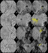
WF-OCTA and en face WF-OCTA images of healthy eyes with different types of PVVs. (A–C) PVV located around the optic disc in a healthy eye. (A) Detection of PVV around the optic disc using 24 × 20 mm en face WF-OCTA in the large-vessel choroidal layer. (B) WF-OCTA image showing the large-vessel choroidal layer in the same scan as A. (C) The yellow highlight indicates the presence of PVV around the optic disc in B. (D–F) PVV located at the macula area in another healthy eye. (D) Detection of PVV at the macula area using 24 × 20 mm en face WF-OCTA in the large-vessel choroidal layer. (E) WF-OCTA image showing the large-vessel choroidal layer in the same scan as D. (F) The yellow highlight indicates the presence of PVV at the macula area in E. (G–I) PVV located at other areas of the posterior pole in another healthy eye. (G) Detection of PVV at other areas of the posterior pole using 24 × 20 mm en face WF-OCTA in the large-vessel choroidal layer. (H) WF-OCTA image showing the large-vessel choroidal layer in the same scan as G. (I) The yellow highlight indicates the presence of PVV at other areas of the posterior pole in H.
Figure 3.
Figure 3.
WF-OCTA and en face WF-OCTA images of both eyes of healthy subjects with PVVs. (A–F) PVV located around the optic disc in both eyes of a healthy subject. (A–C) PVV located around the optic disc in the right eye. (A) Detection of PVV around the optic disc in the right eye using 24 × 20 mm en face WF-OCTA in the large-vessel choroidal layer. (B) WF-OCTA image showing the large-vessel choroidal layer in the same scan as A. (C) The yellow highlight indicates the presence of PVV around the optic disc in B. (D–F) PVV located around the optic disc in the left eye. (D) Detection of PVV around the optic disc in the left eye using 24 × 20 mm en face WF-OCTA in the large-vessel choroidal layer. (E) WF-OCTA image showing the large-vessel choroidal layer in the same scan as D. (F) The yellow highlight indicates the presence of PVV around the optic disc in E. (G–L) PVV located at other areas of the posterior pole in both eyes of another healthy subject. (G–I) PVV located at other areas of the posterior pole in the right eye. (G) Detection of PVV at other areas of the posterior pole in the right eye using 24 × 20 mm en face WF-OCTA in the large-vessel choroidal layer. (H) WF-OCTA image showing the large-vessel choroidal layer in the same scan as G. (I) The yellow highlight indicating the presence of PVV at other areas of the posterior pole in H. (J–L) PVV located at other areas of the posterior pole in the left eye. (J) Detection of PVV at other areas of the posterior pole in the left eye using 24 × 20 mm en face WF-OCTA in the large-vessel choroidal layer. (K) WF-OCTA image showing the large-vessel choroidal layer in the same scan as J. (L) The yellow highlight indicates the presence of PVV at other areas of the posterior pole in K.
Figure 4.
Figure 4.
WF-OCTA and en face WF-OCTA images of a single eye of healthy subjects with two types of PVVs. (A–C) PVVs located around the optic disc and at other areas of the posterior pole in a single eye of a healthy subject. (A) Detection of two types of PVVs in the healthy eye using 24 × 20 mm en face WF-OCTA in the large-vessel choroidal layer. (B) WF-OCTA image showing the large-vessel choroidal layer in the same scan as A. (C) The yellow highlight indicating the presence of two types of PVVs in B. (D–F) PVVs located around the optic disc and at other areas of the posterior pole in a single eye of another healthy subject. (D) Detection of two types of PVVs in the healthy eye using 24 × 20 mm en face WF-OCTA in the large-vessel choroidal layer. (E) WF-OCTA image showing the large-vessel choroidal layer in the same scan as D. (F) The yellow highlight indicates the presence of two types of PVVs in E.
Figure 5.
Figure 5.
WF-OCTA and en face WF-OCTA images of a single eye of healthy subjects with multiple PVVs. (A–C) Two PVVs located around the optic disc in a single eye of a healthy subject. (A) Detection of two PVVs around the optic disc in the healthy eye using 24 × 20 mm en face WF-OCTA in the large-vessel choroidal layer. (B) WF-OCTA image showing the large-vessel choroidal layer in the same scan as A. (C) The yellow highlight indicates the presence of two PVVs around the optic disc in B. (D–F) Six PVVs located around the optic disc in a single eye of another healthy subject. (D) Detection of six PVVs around the optic disc in the healthy eye using 24 × 20 mm en face WF-OCTA in the large-vessel choroidal layer. (E) WF-OCTA image showing the large-vessel choroidal layer in the same scan as D. (F) The yellow highlight indicates the presence of six PVVs around the optic disc in E. (G–I) Two PVVs located around the optic disc in a single healthy eye of another healthy subject. (G) Detection of two PVVs around the optic disc in the healthy eye using 24 × 20 mm en face WF-OCTA in the large-vessel choroidal layer. (H) WF-OCTA image showing the large-vessel choroidal layer in the same scan as G. (I) The yellow highlight indicates the presence of two PVVs around the optic disc in H.

Similar articles

Cited by

References

    1. Hayreh SS, Hayreh SB.. Uveal vascular bed in health and disease: uveal vascular bed anatomy. Paper 1 of 2. Eye 2023. Available at: https://www.nature.com/articles/s41433-023-02416-z. Accessed May 22, 2023. - PMC - PubMed
    1. Nickla DL, Wallman J.. The multifunctional choroid. Prog Retinal Eye Res. 2010; 29: 144–168. - PMC - PubMed
    1. Yu D-Y, Yu PK, Cringle SJ, et al. .. Functional and morphological characteristics of the retinal and choroidal vasculature. Prog Retinal Eye Res. 2014; 40: 53–93. - PubMed
    1. Verma A, Maram J, Alagorie AR, et al. .. Distribution and location of vortex vein ampullae in healthy human eyes as assessed by ultra-widefield indocyanine green angiography. Ophthalmol Retina. 2020; 4: 530–534. - PubMed
    1. Lim MC, Bateman JB, Glasgow BJ.. Vortex vein exit sites. Ophthalmology. 1995; 102: 942–946. - PubMed