Review
. 2024 Feb 9;24(1):63.
doi: 10.1186/s12935-024-03220-9.

Melanoma biology and treatment: a review of novel regulated cell death-based approaches

Affiliations
Review

Melanoma biology and treatment: a review of novel regulated cell death-based approaches

Ming-Yun Hsieh et al. Cancer Cell Int. .

Abstract

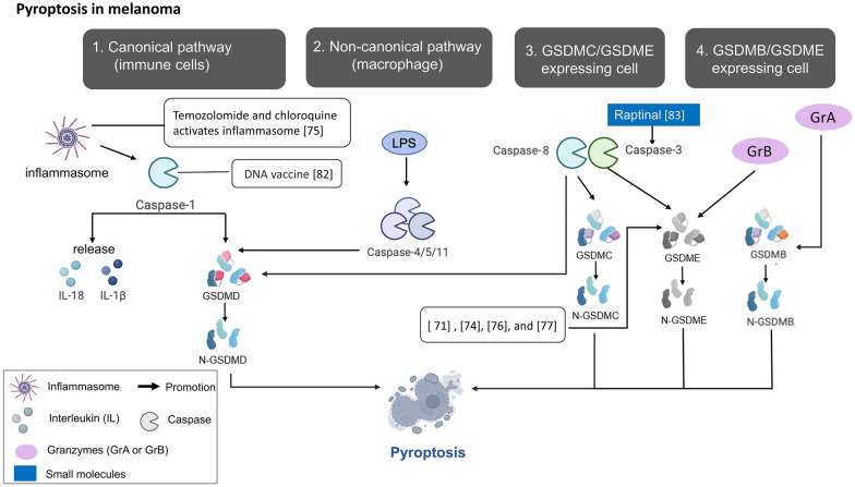
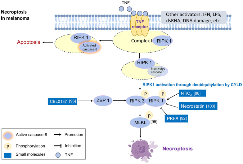
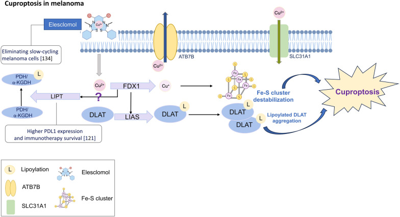
The incidence of melanoma, the most lethal form of skin cancer, has increased due to ultraviolet exposure. The treatment of advanced melanoma, particularly metastatic cases, remains challenging with poor outcomes. Targeted therapies involving BRAF/MEK inhibitors and immunotherapy based on anti-PD1/anti-CTLA4 antibodies have achieved long-term survival rates of approximately 50% for patients with advanced melanoma. However, therapy resistance and inadequate treatment response continue to hinder further breakthroughs in treatments that increase survival rates. This review provides an introduction to the molecular-level pathogenesis of melanoma and offers an overview of current treatment options and their limitations. Cells can die by either accidental or regulated cell death (RCD). RCD is an orderly cell death controlled by a variety of macromolecules to maintain the stability of the internal environment. Since the uncontrolled proliferation of tumor cells requires evasion of RCD programs, inducing the RCD of melanoma cells may be a treatment strategy. This review summarizes studies on various types of nonapoptotic RCDs, such as autophagy-dependent cell death, necroptosis, ferroptosis, pyroptosis, and the recently discovered cuproptosis, in the context of melanoma. The relationships between these RCDs and melanoma are examined, and the interplay between these RCDs and immunotherapy or targeted therapy in patients with melanoma is discussed. Given the findings demonstrating melanoma cell death in response to different stimuli associated with these RCDs, the induction of RCD shows promise as an integral component of treatment strategies for melanoma.

Keywords: Autophagy-dependent cell death; Cuproptosis; Ferroptosis; Immunotherapy; Melanoma; Necroptosis; Pyroptosis; Regulated cell death; Targeted therapy.


Conflict of interest statement

No competing interests.

Figures

Fig. 1
Fig. 1
Schematic diagram of autophagy-dependent cell death and related research in melanoma. Autophagy is a process initiated by ULK1 complex formation, followed by membrane isolation and phagospore formation. After fusion with lysosome, autophagosome ends up and engulfed substrates are digested in autophagosome. Related researches are expressed in white squares with reference number in parenthesis. The exogenous small molecules are marked in blue squares especially. For example, vemurafenib (a BRAF inhibitor) resistance is associated with elevated expression of Beclin-1, ATG5 and UVRAG, and its efficacy can be increased by ectopic expression of miR-216b to inhibit ATG5. The manipulation of autophagy can also influence the immune response in melanoma, such as increased local infiltration of NK cells in melanoma after inhibition of Beclin-1, ATG5 or p62 correlated with increased expression of CCL5 and improved prognosis. Loss of BNIP3 decreased macrophage phagocytosis of dying melanoma cells. Besides, autophagy activity is associated with MDSC- mediated suppression of anti-melanoma immunity, melanoma adaption to fluctuating O2 or pH, and outcome of targeted therapy with temozolomide and sorafenib
Fig. 2
Fig. 2
Pyroptosis and its role in melanoma. Pyroptosis can be activated by four pathways: (1) the canonical pathway with inflammasome assembly and caspase-1 activation (2) the noncanonical pathway with caspase-4/5/11 activation by direct binding of its N-terminal CARD domain to intracellular LPS. (3) Caspase 3/8-mediated pathway with GSDMC or GSDME cleavage (4) Granzyme-mediated pathway with granzyme A and B, secreted from cytotoxic T lymphocytes and NK cells respectively, can cleave GSDMB and GSDME, respectively. Research of pyroptosis on melanoma was expressed in white squares with reference number in parenthesis. The exogenous molecules are marked in blue squares especially. GSDME cleavage by activated caspase 3 has been observed by chemotherapeutic drugs, BRAF and MEK inhibitors/ PDPK1 and MEK inhibitors, and ROS production with iron dextran or raptinal, a caspase 3 activator. Caspase-1 DNA was ever included in anticancer DNA vaccine to induce pyroptosis of melanoma cells. Pyroptosis can also be induced by temozolomide and chloroquine via autophagy inhibition and inflammasome activation
Fig. 3
Fig. 3
Schematic diagram of necroptosis and its effects on melanoma. Necroptosis can be triggered by activation of death receptors or Toll-like receptors. Through interaction of activated RIPK1 with RIPK3, and subsequently, RIPK3 phosphorylates MLKL. Pore formation on cell membrane is the final step of necroptosis. Research of necroptosis on melanoma was expressed in white squares with reference number in parenthesis. The exogenous molecules are marked in blue squares especially. While unsure effect of RIPK1 inhibition on melanoma metastasis, direct intratumor delivery of MLKL mRNA can inhibit melanoma tumor growth and metastasis and augment the efficiency of immune blockade therapy. Besides, NTiO2, BAY 87–2243 and CBL0137 are the agents with the ability to induce necroptosis in melanoma cells
Fig. 4
Fig. 4
The relationship between ferroptosis and melanoma. Ferroptosis is characterized by the overwhelming production of ROS and accumulation of iron-dependent lipid peroxides. Research of ferroptosis on melnoma was expressed in white squares with reference number in parenthesis. The exogenous molecules are marked in blue squares especially. Ferroptosis can be induced in melanoma cells by GPX4 inhibition in the situation of BRAF inhibitor resistance or with the application of plant-derived phyto-sesquiterpene lactone. Nobiletin can also induce ferroptosis in melanoma cells through another pathway of GSK3β-mediated Keap1/Nrf2/HO-1 signaling. In addition, IFNγ release after immunotherapy can drive ferroptosis by downregulating the expression of system Xc. which can be further enhanced by the delivery of miR-21-3p-loaded gold nanoparticles
Fig. 5
Fig. 5
Schematic diagram of cuproptosis and correlation between cuproptosis and melanoma. Copper ions can cross the cell membrane into the intracellular space with a copper ionophore, and their concentration can be regulated by copper importers/exporters (SLC31A1/ATP7A and B). FDX1 not only reduces divalent copper to more toxic monovalent copper but also regulates protein lipoylation. Intracellular copper can bind lipoylated proteins directly, leading to their aggregation and further occurrence of cuproptosis. Copper ions also contribute to Fe-S cluster loss, which is another mechanism of cuproptosis. Related researches are expressed in white squares with reference number in parenthesis. Elesclomol, a copper ionophore, can induce cuproptosis, and was shown to eliminate slow-cycling melanoma cells. A higher expression level of LIPT1, responsible for transferring lipoic acid to the E2 subunits of AKGDH and pyruvate dehydrogenase PDH, is related to longer survival after immunotherapy

References

    1. Chang AE, Karnell LH, Menck HR. The National Cancer Data Base report on cutaneous and noncutaneous melanoma: a summary of 84,836 cases from the past decade. Cancer. 1998;83(8):1664–1678. doi: 10.1002/(SICI)1097-0142(19981015)83:8<1664::AID-CNCR23>3.0.CO;2-G. - DOI - PubMed
    1. Siegel RL, Miller KD, Wagle NS, Jemal A. Cancer statistics, 2023. CA Cancer J Clin. 2023;73(1):17–48. doi: 10.3322/caac.21763. - DOI - PubMed
    1. Klemen ND, Wang M, Rubinstein JC, Olino K, Clune J, Ariyan S, Cha C, Weiss SA, Kluger HM, Sznol M. Survival after checkpoint inhibitors for metastatic acral, mucosal and uveal melanoma. J Immunother Cancer. 2020;8(1):e000341. doi: 10.1136/jitc-2019-000341. - DOI - PMC - PubMed
    1. McLaughlin CC, Wu XC, Jemal A, Martin HJ, Roche LM, Chen VW. Incidence of noncutaneous melanomas in the US. Cancer. 2005;103(5):1000–1007. doi: 10.1002/cncr.20866. - DOI - PubMed
    1. Schadendorf D, Fisher DE, Garbe C, Gershenwald JE, Grob JJ, Halpern A, Herlyn M, Marchetti MA, McArthur G, Ribas A, et al. Melanoma. Nat Rev Dis Primers. 2015;1:15003. doi: 10.1038/nrdp.2015.3. - DOI - PubMed