. 2024 Feb 9;22(1):56.
doi: 10.1186/s12951-024-02330-w.

M2 microglia-derived exosomes promote vascular remodeling in diabetic retinopathy

Affiliations

M2 microglia-derived exosomes promote vascular remodeling in diabetic retinopathy

Xingxing Wang et al. J Nanobiotechnology. .

Abstract

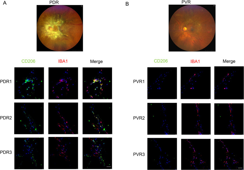
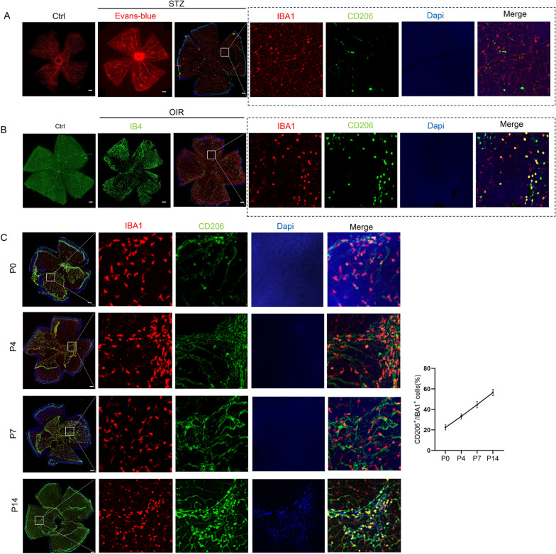
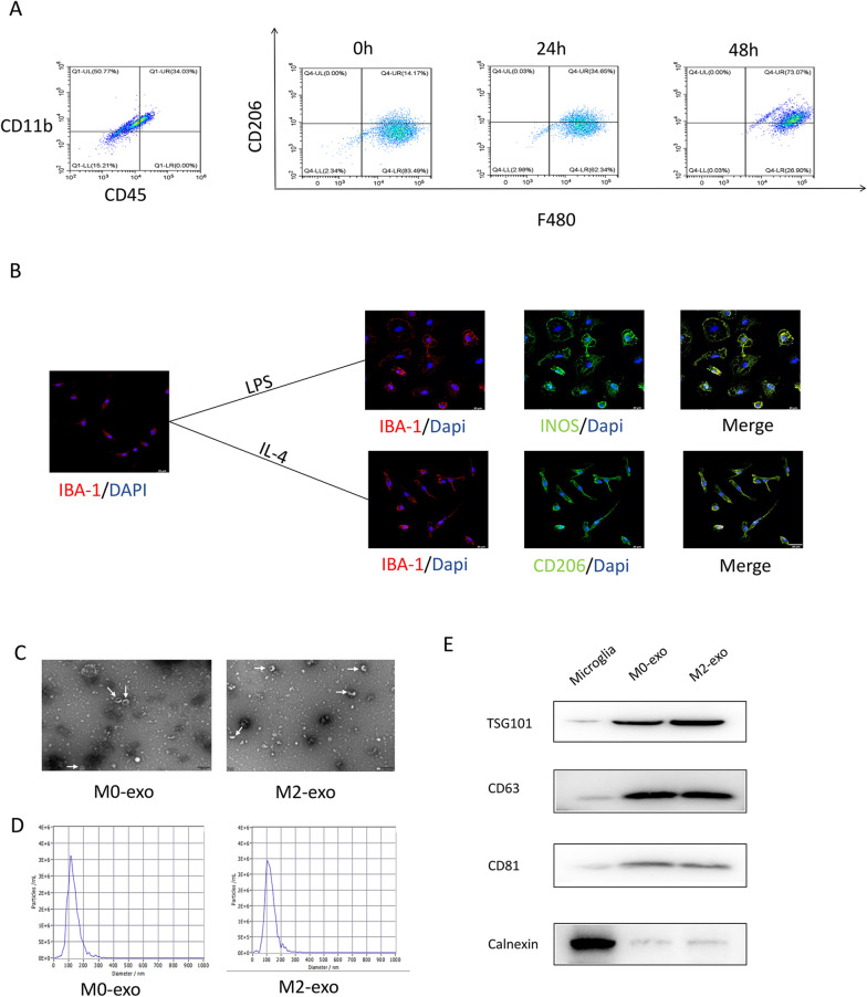
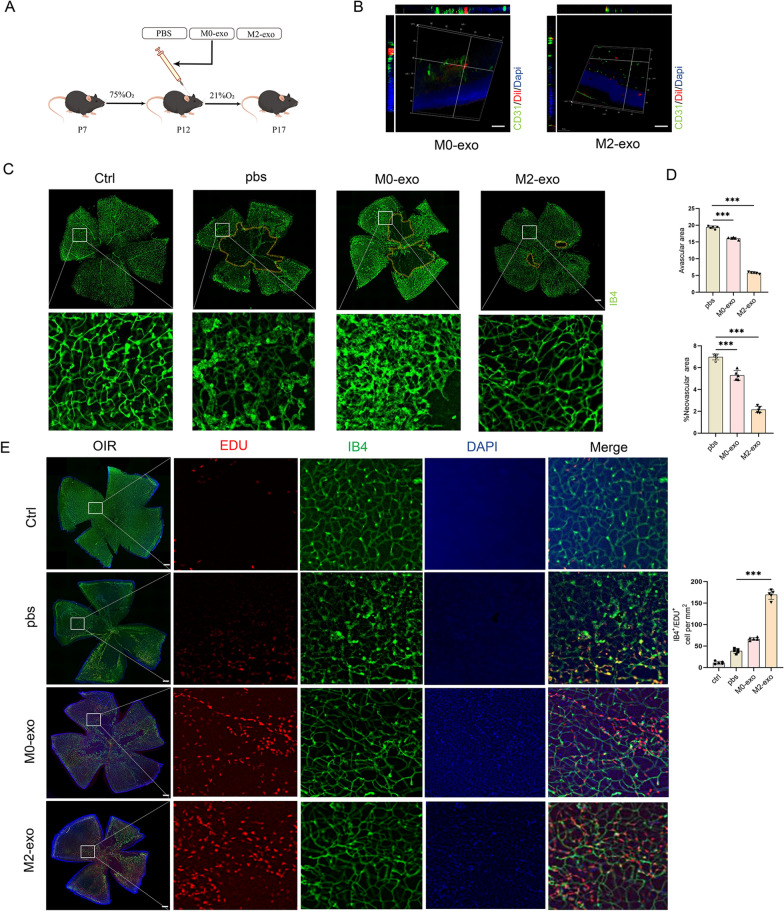
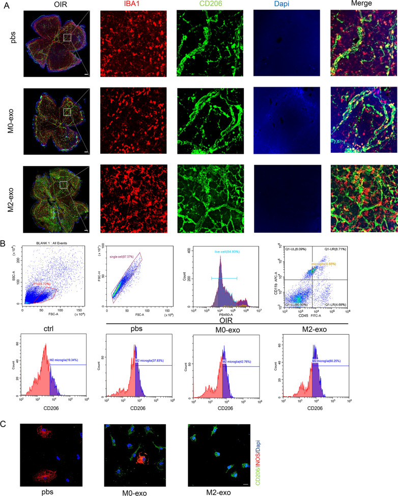
Diabetic retinopathy (DR) is a vision-threatening diabetic complication that is characterized by microvasculature impairment and immune dysfunction. The present study demonstrated that M2 microglia intensively participated in retinal microangiopathy in human diabetic proliferative membranes, mice retinas, retinas of mice with oxygen-induced retinopathy (OIR) mice, and retinas of streptozotocin-induced DR mice. Further in vivo and in vitro experiments showed that exosomes derived from M2 polarized microglia (M2-exo) could reduce pericyte apoptosis and promote endothelial cell proliferation, thereby promoting vascular remodeling and reducing vascular leakage from the diabetic retina. These effects were further enhanced by M2-exo that facilitated M2 polarization of retinal microglia. Collectively, the study demonstrated the capability of M2-exo to induce retinal microvascular remodeling, which may provide a new therapeutic strategy for the treatment of DR.


Conflict of interest statement

No potential conflicts of interest relevant to this article were reported.

Figures

Fig. 1
Fig. 1
Immunohistochemical staining of M2 microglia in the epiretinal fibrovascular membrane obtained from patients with PDR (A) and PVR (B) (scale bar = 50 μm)
Fig. 2
Fig. 2
Role of M2 microglia in OIR and STZ-induced DR mouse models (scale bar = 200 μm). A Involvement of M2 microglia in retinal vascular changes in STZ-induced DR mouse model. B Involvement of M2 microglia in retinal vascular changes in OIR mouse model. C Involvement of M2 microglia in normal blood vessel formation
Fig. 3
Fig. 3
Identification of M2 microglia and exosomes. A Microglia were induced with IL-4 for 0, 24, and 48 h for flow identification. B Identification of primary microglial polarization by immunofluorescence (scale bar = 20 μm). C TEM images of exosome ultrastructure (white arrows represent exosomes). D Nanoparticle tracking analysis of M0 and M2 exosomes. E Representative bands showing protein levels of the exosomal surface markers TSG101, CD63, CD81, and calnexin
Fig. 4
Fig. 4
Promotion of in vivo retinal angiogenesis and remodeling by exosomes of M2 microglia. A Flow chart describes the in vivo establishment of the OIR mouse model. B Three-dimensional view of a frozen retinal section of OIR mouse showing exosome uptake in vivo. Scale bar = 50 μm. C, D Retinal flat mounts of C57BL/6J mice after OIR induction at P17 and intravitreal injection of M0-exo, M2-exo, or PBS at P12; yellow indicates the avascular area. Compared with the PBS or M0-exo group, the M2-exo group demonstrated significantly reduced avascular region and abnormal proliferating vessels. *p < 0.05, **p < 0.01***p < 0.001; n = 5. Scale bar = 200 μm. E Retinal flat mounts after tail vein injection of EdU in OIR mice. Effects of M2-exo on cell proliferation are demonstrated as the highest EdU+ cell number. ***p < 0.001; n = 5. Scale bar = 200 μm
Fig. 5
Fig. 5
M2-exo attenuates retinal vascular leakage and reduces the number of acellular capillaries. A Three-dimensional view of a frozen retinal section of mice with STZ-induced DR showing exosome uptake in vivo. Scale bar = 100 μm. B, C E/P ratio, number of acellular strands, and morphological changes in vessels among the four groups assessed using PAS staining of retinal tissue (*p < 0.05, **p < 0.01). White arrows indicate acellular strands. n = 5. Scale bar = 50 μm. E/P endothelial cell-to-pericyte. D, E Microvascular permeability of whole retinal branches evaluated by Evans blue fluorescence staining. Dye leakage was observed outwards from the retinal vessels in the STZ-induced DR mouse model, whereas almost no Evans blue leakage was observed in the M2-exo-treated group. *p < 0.05, **p < 0.01***p < 0.001; n = 5. Scale bar = 200 μm. F Quantification of Evans blue content in the retina. ***p < 0.001; n = 5
Fig. 6
Fig. 6
Effect of exosomes from M2 microglia on hRECs. A The uptake of hRECs into exosomes was detected by labeling them with Dil. Scale bar = 20 μm. B Tube formation ability was measured. Migration of hRECs was determined by Transwell assay. Scale bar = 200 μm. C, D Proliferation of hRECs was assessed by the EdU assay. The percentage of EdU-positive cells (marked in red) was calculated using three randomly selected regions. Scale bar = 20 μm. E, F Immunofluorescence detection and quantification of ZO-1 in hRECs. Scale bar = 20 μm. G The morphology of vascular TJs in each group of mice was observed by TEM (arrows represent TJs). Scale bar = 2 μm
Fig. 7
Fig. 7
Effect of M2 microglia-derived exosomes on pericytes. A Identification of pericyte fluorescent staining (scale bar = 20 μm) and pericyte morphology using light microscopy. Scale bar = 200 μm. B Uptake of M2 exosomes by pericytes. Scale bar = 20μm. C, D Flow cytometry analysis of pericytes for apoptosis and quantification. E, F Blood–retinal barrier integrity was assessed by transendothelial electrical resistance (TEER) and fluorescein isothiocyanate (FITC)-dextran in co-cultures of hRECs with pericytes
Fig. 8
Fig. 8
M2-exo promotes revascularization and facilitates M2 polarization of microglia. A Validation of the ability of M2-exo to promote revascularization and M2 microglia polarization by immunofluorescence in OIR mouse (scale bar = 200 μm). B Retinal cell flow analysis demonstrates that M2-exo promotes M2-type microglial polarization. C Representative immunofluorescence images showing changes in primary microglia after treatment with PBS, M0-exo, and M2-exo. Scale bar = 20 μm

References

    1. Yau JW, Rogers SL, Kawasaki R, Lamoureux EL, Kowalski JW, Bek T, Chen SJ, Dekker JM, Fletcher A, Grauslund J, et al. Global prevalence and major risk factors of diabetic retinopathy. Diabetes Care. 2012;35(3):556–564. doi: 10.2337/dc11-1909. - DOI - PMC - PubMed
    1. Altmann C, Schmidt MHH. The role of microglia in diabetic retinopathy: inflammation, microvasculature defects and neurodegeneration. Int J Mol Sci. 2018;19(1):110. doi: 10.3390/ijms19010110. - DOI - PMC - PubMed
    1. Armulik A, Abramsson A, Betsholtz C. Endothelial/pericyte interactions. Circ Res. 2005;97(6):512–523. doi: 10.1161/01.RES.0000182903.16652.d7. - DOI - PubMed
    1. Potente M, Gerhardt H, Carmeliet P. Basic and therapeutic aspects of angiogenesis. Cell. 2011;146(6):873–887. doi: 10.1016/j.cell.2011.08.039. - DOI - PubMed
    1. Boss JD, Singh PK, Pandya HK, Tosi J, Kim C, Tewari A, Juzych MS, Abrams GW, Kumar A. Assessment of neurotrophins and inflammatory mediators in vitreous of patients with diabetic retinopathy. Invest Ophthalmol Vis Sci. 2017;58(12):5594–5603. doi: 10.1167/iovs.17-21973. - DOI - PMC - PubMed